Where are heart surgeries performed? The main types of surgical intervention on the heart. Action plan before surgery
Open heart surgery is one of the methods of treating cardiovascular diseases, in which special surgical procedures are performed. The general principle is that there is an intervention in the human body in order to carry out the necessary activities on the open heart. In other words, this is such an operation, during which an opening or dissection of the region of the human sternum is performed, affecting the tissues of the organ itself and its vessels.
Open heart surgery
Statistics show that the most common intervention of this type among adults is an operation in which artificial blood flow is created from the aorta to healthy areas of the coronary arteries - coronary artery bypass grafting.
This operation is performed for the treatment of severe coronary heart disease, which occurs due to the development of atherosclerosis, in which there is a narrowing of the vessels supplying blood to the myocardium, their elasticity decreases.
The general principle of the operation: the patient's own biomaterial (a fragment of an artery or vein) is taken and sutured in the area between the aorta and the coronary vessel in order to bypass the place affected by atherosclerosis, in which blood circulation is impaired. After the operation is performed, the blood supply to a certain area of the heart muscle is restored. This artery / vein supplies the heart with the necessary blood flow, while the artery in which the pathological process occurs is bypassed.
 Coronary artery bypass grafting
 Today, taking into account the progress in medicine, for surgical treatment on the heart, it is enough to make only small incisions in the corresponding area. Another intervention, more complex, will not be needed. Therefore, the concept of "open heart surgery" sometimes misleads people.
Reasons for open heart surgery
- The need to replace or restore the patency of blood vessels for the correct flow of blood into the heart.
- The need to repair defective areas in the heart (for example, valves).
- The need to place special medical devices to maintain the working capacity of the heart.
- The need for transplantation operations.
What you need to know about coronary artery bypass surgery?
Time spending
According to medical data, this type of operation takes no less than four and no more than six hours. In rare, especially severe cases, when the operation requires more work (creation of several shunts), an increase in this period may be observed.
The first night after heart surgery and all medical procedures, patients spend in the intensive care unit. After three to seven days have passed (the exact number of days is determined by the patient's well-being), the person is transferred to a regular ward.
Operation Hazards
Despite the qualifications of doctors, no one is immune from unplanned situations. What is the danger of surgery, and what risk can it carry:
- infection of the chest due to an incision (this risk is especially high for people who are obese, diabetic, or undergo a second operation);
- myocardial infarction, ischemic stroke;
- heart rhythm disturbances;
- thromboembolism;
- increased body temperature for a long time;
- cardiac discomfort of any nature;
- pain of a different nature in the chest area;
- pulmonary edema;
- short-term amnesia and other transient memory problems;
- loss of a significant amount of blood.
These negative consequences, as statistics show, occur much more often when using an artificial blood supply device.
 The risk of unpleasant consequences is always present
 Preparation period
In order for the planned operation and general treatment to be successful, it is important not to miss anything significant before they begin. To do this, the patient must tell the doctor:
- About medications that are currently being used. These may include medications prescribed by another doctor, or those that the patient purchases himself, including dietary supplements, vitamins, etc. This is important information and should be announced before surgery.
- About all chronic and past diseases, health deviations that are currently available (runny nose, herpes on the lips, indigestion, fever, sore throat, fluctuations in blood pressure, etc.).
The patient should be prepared for the fact that two weeks before the operation, the doctor will ask him to refrain from smoking, excessive alcohol consumption, taking vasoconstrictor drugs (for example, nasal drops, ibuprofen, etc.).
On the day of the operation, the patient will be asked to use a special antibacterial soap, which significantly reduces the risk of infection during the operation. In addition, a few hours before the intervention, you can not eat food and drink water.
Operation
When open heart surgery is performed, the following actions are sequentially performed:
- The patient is placed on the operating table.
- He is given general anesthesia.
- When the anesthesia begins to take effect and the patient falls asleep, the doctor opens the chest. To do this, he makes an incision in the appropriate area (usually it is no more than 25 sentiments in length).
- The doctor dissects the sternum, partially or completely. This allows access to the heart and aorta.
- Once access is secured, the patient's heart is stopped and connected to a heart-lung machine. This allows the surgeon to calmly perform all manipulations. Today, technologies are used that allow in some cases to perform this operation without stopping the heartbeat, while the number of complications is lower. than traditional intervention.
- The doctor creates a shunt around the damaged part of the artery.
- The cut part of the chest is fixed with a special material, most often with a special wire, but in some cases plates are used. These plates are often used for the elderly or for people who have had frequent surgeries.
- After the operation is completed, the incision is sutured.
Postoperative period
After the operation is completed and the patient wakes up, he will find two or three tubes in his chest. The role of these tubes is to drain excess fluid from the area around the heart (drainage) into a special vessel. In addition, an intravenous tube is installed for the flow of therapeutic and nutrient solutions into the body and a catheter into the bladder to remove urine. In addition to the tubes, devices are connected to the patient to monitor the work of the heart.
The patient should not worry, in case of questions or discomfort, he can always contact the medical workers who will be assigned to monitor him and promptly respond if necessary.
 The duration of the recovery period depends not only on physiology, but also on the person himself.
 Each patient should understand that rehabilitation after surgery is not a quick process. After six weeks of treatment, some improvements can be observed, and only after six months will all the benefits of the operation become visible.
But each patient is able to speed up this rehabilitation process, while avoiding new heart ailments, which reduces the risk of a second operation. To do this, it is recommended to take the following measures:
- follow the diet and special diet prescribed by the attending physician;
- limit salty, fatty, sweet foods);
- devote time to physiotherapy exercises, walks in the fresh air;
- stop drinking alcohol frequently;
- monitor the level of cholesterol in the blood;
- track blood pressure.
If these measures are followed, the postoperative period will pass quickly and without complications. But you should not rely on general recommendations, the advice of your attending physician, who has studied the medical history in detail and is able to draw up an action plan and a diet during the recovery period, is much more valuable.
Cardiac surgery is a branch of medicine dedicated to the surgical treatment of the heart. With pathologies of the cardiovascular system, such intervention is an extreme measure. Doctors try to restore the patient's health without surgery, but in some cases only cardiac surgery can save the patient. Today, this field of cardiology uses the latest advances in science to return the patient to health and a fulfilling life.
Indications for operations
Invasive interventions on the heart is a complex and risky job, it requires skill and experience, and the patient - preparation and implementation of recommendations. Since such operations are risky, they are carried out only when absolutely necessary. In most cases, the patient is trying to rehabilitate with the help of medicines and medical procedures. But in cases where such methods do not help, heart surgery is needed. Surgical intervention is carried out in a hospital and complete sterility, the operated is under anesthesia and the control of the surgical team.
Such interventions are needed for congenital heart defects or acquired. The former include pathologies in the anatomy of the organ: defects in valves, ventricles, impaired blood circulation. Most often they are discovered even during the bearing of a child. Heart disease is also diagnosed in newborns, often such pathologies need to be eliminated urgently in order to save the life of the baby. Among the acquired diseases, ischemic disease is in the lead, in this case, surgery is considered the most effective method of treatment. Also in the heart area there are: impaired blood circulation, stenosis or valve insufficiency, heart attack, pericardial pathology and others.
Heart surgery is prescribed in situations where conservative treatment does not help the patient, the disease progresses rapidly and threatens life, with pathologies that require urgent and urgent correction, and in advanced forms of diseases, a late visit to the doctor.
The decision on the appointment of the operation is made by a council of doctors or. The patient must be examined to establish an accurate diagnosis and type of surgical intervention. They identify chronic diseases, stages of the disease, assess the risks, in which case they talk about a planned operation. If emergency assistance is needed, for example, when a blood clot is torn off or an aneurysm is exfoliated, minimal diagnostics are performed. In any case, the function of the heart is restored surgically, its departments are rehabilitated, blood flow and rhythm are normalized. In severe situations, the organ or its parts are no longer amenable to correction, then prosthetics or transplantation is prescribed.
Classification of heart operations
 In the area of the heart muscle, there can be dozens of different diseases, these are: insufficiency, narrowing of the lumen, ruptures of blood vessels, stretching of the ventricles or atria, purulent formations in the pericardium, and much more. To solve each problem, surgery has several types of operations. They are distinguished by urgency, effectiveness and method of influencing the heart.
The general classification divides them into operations:
- Buried - used to treat arteries, large vessels, aorta. During such interventions, the chest of the operated person is not opened, the heart itself is also not affected by the surgeon. Therefore, they are called "closed" - the heart muscle remains intact. Instead of a strip opening, the doctor makes a small incision in the chest, most often between the ribs. Closed types include: shunting, balloon angioplasty, stenosis of blood vessels. All these manipulations are designed to restore blood circulation, sometimes they are prescribed to prepare for a future open operation.
- Open - carried out after opening the sternum, sawing the bones. The heart itself during such manipulations can also be opened to get to the problem area. As a rule, for such operations, the heart and lungs must be stopped. To do this, connect the heart-lung machine - AIC, it compensates for the work of "disabled" organs. This allows the surgeon to accurately perform the work, in addition, the procedure under the control of AIC takes longer, which is necessary when eliminating complex pathologies. During open operations, AIC may not be connected, but only the desired zone of the heart can be stopped, for example, during coronary artery bypass grafting. Opening the chest is necessary to replace valves, prosthetics, and eliminate tumors.
- X-ray surgery - similar to a closed type of operation. The essence of this method is that the doctor moves a thin catheter through the blood vessels, and gets to the very heart. The chest is not opened, the catheter is placed in the thigh or shoulder. The catheter is injected with a contrast agent that stains the vessels. The catheter is advanced under X-ray control, the video image is transmitted to the monitor. Using this method, the lumen in the vessels is restored: at the end of the catheter there is a so-called balloon and a stent. At the site of narrowing, this balloon is inflated with a stent, restoring the normal patency of the vessel.
The safest are minimally invasive methods, that is, X-ray surgery and a closed type of surgery. With such work, the risk of complications is the least, the patient recovers faster after them, but they can not always help the patient. Complex operations can be avoided with periodic inspections. The earlier the problem is identified, the easier it is for the doctor to solve it.
 Depending on the condition of the patient, there are:
- planned operation. It is carried out after a detailed examination, within the agreed time frame. A planned intervention is prescribed when the pathology does not pose a particular danger, but it cannot be postponed.
- Urgent - these are operations that need to be done in the next few days. During this time, the patient is prepared, all the necessary studies are carried out. The date is set immediately after receiving the necessary data.
- Emergency. If the patient is already in serious condition, the situation may worsen at any time - an operation is prescribed immediately. Before her, only the most important examinations and preparations are carried out.
In addition, surgical care can be radical or auxiliary. The first implies the complete elimination of the problem, the second - the elimination of only part of the disease, improving the patient's well-being. For example, if a patient has a pathology of the mitral valve and stenosis of a vessel, the vessel is first restored (auxiliary), and after a while valve plastic surgery (radical) is prescribed.
How operations are done
The course and duration of the operation depends on the pathology being eliminated, the patient's condition, and the presence of concomitant diseases. The procedure can take half an hour, and can stretch for 8 hours or more. Most often, such interventions last 3 hours, are carried out under general anesthesia and AIC control. First, the patient is prescribed an ultrasound of the chest, urine and blood tests, an ECG, and a consultation with specialists. After receiving all the data, they determine the degree and place of the pathology, decide whether there will be an operation.
As part of the preparation, a low-fat, spicy, and fried diet is also prescribed. For 6-8 hours before the procedure, it is recommended to refuse food and drink less. In the operating room, the doctor assesses the well-being of the ward, introduces the patient into a medical sleep. With minimally invasive interventions, local anesthesia is sufficient, for example, during X-ray surgery. When anesthesia or anesthesia takes effect, the main actions begin.
Heart valve repair
There are four valves in the heart muscle, all of which serve as a passage for blood from one chamber to another. The most commonly operated valves are the mitral and tricuspid valves, which connect the ventricles to the atria. Stenosis of the passages occurs with insufficient expansion of the valves, while the blood does not flow well from one department to another. Valve insufficiency is a poor closure of the cusps of the passage, while there is an outflow of blood back.
 Plastic surgery is carried out open or closed, during the operation, special rings or sutures are applied manually along the diameter of the valve, which restore the normal lumen and narrow the passage. Manipulations last an average of 3 hours; with open views, an AIC is connected. After the procedure, the patient remains under the supervision of doctors for at least a week. The result is normal blood circulation and functioning of the heart valves. In severe cases, native leaflets are replaced with artificial or biological implants.
Elimination of heart defects
In most cases, defects are congenital, the cause of this can be hereditary pathologies, bad habits of parents, infections and fever during pregnancy. At the same time, children may have various anatomical abnormalities in the region of the heart, often such anomalies are poorly compatible with life. The urgency and type of surgery depends on the condition of the child, but they are often prescribed as early as possible. For children, heart surgery is performed only under general anesthesia, and under the supervision of medical equipment.
At an older age, heart defects develop with defects in the interatrial septum. This happens with mechanical damage to the chest, infectious diseases, due to concomitant heart disease. To eliminate such a problem, an open operation is also needed, more often with artificial cardiac arrest.
During manipulations, the surgeon can “patch” the septum with a patch, or suture the defective part.
Shunting
Coronary artery disease (CHD) is a very common pathology that affects mainly the generation over 50 years of age. Appears due to impaired blood flow in the coronary artery, which leads to oxygen starvation of the myocardium. There is a chronic form, in which the patient has constant attacks of angina pectoris, and an acute one is a myocardial infarction. They try to eliminate chronic pain conservatively or with the help of minimally invasive techniques. Acute requires urgent intervention.
To prevent complications or alleviate the disease, apply:
- aorto-coronary bypass;
- balloon angioplasty;
- transmyocardial laser revascularization;
- stenting of a coronary artery.
 All these methods are aimed at restoring normal blood flow. As a result, enough oxygen is supplied to the myocardium with blood, the risk of a heart attack is reduced, and angina pectoris is eliminated.
If you need to restore normal patency, angioplasty or stenting is enough, in which the catheter is moved through the vessels to the heart. Before such an intervention, coronary angiography is performed to accurately determine the blocked area. Sometimes blood flow is restored bypassing the affected area, while a bio-shunt (often a section of the patient's own vein from the arm or leg) is sutured to the artery.
Recovery after interventions
After surgery, the patient remains in the hospital for another 1-3 weeks, all this time the doctors will assess his condition. The patient is discharged after verification and approval by the cardiologist.
The first month after surgical procedures is called the early postoperative period, at this time it is very important to follow all the doctor's recommendations: diet, calm and measured lifestyle. Nicotine, alcohol, junk food and physical activity are prohibited regardless of the type of intervention.
The doctor's recommendations should also contain a warning about the dangers and complications. At discharge, the doctor will set the date for the next appointment, but you need to seek help and unscheduled if the following symptoms occur:
- sudden fever;
- redness and swelling at the incision site;
- discharge from the wound;
- persistent chest pain;
- frequent dizziness;
- nausea, bloating and stool disorders;
- breathing difficulties.
At scheduled examinations, the cardiologist will listen to the heartbeat, measure the pressure, and listen to complaints. To check the effectiveness of the operation, ultrasound, computed tomography, x-ray examinations are prescribed. Such visits are scheduled once a month for six months, then the doctor will see you once every 6 months.
Often, in addition to surgical care, medications are prescribed. For example, when prosthetic valves are artificially implanted, the patient drinks anticoagulants for life.
In the postoperative period, it is important not to self-medicate, since the interaction of permanent drugs and other medications can give a negative result. Even conventional painkillers need to be discussed with. To keep fit and restore health faster, it is recommended to be outdoors more often, walk on foot.
Life after heart surgery will gradually return to its previous course, a full recovery is predicted within a year.
Cardiac surgery offers many methods for the rehabilitation of the heart. Such operations are designed to restore the patient's physical and moral strength. You should not be afraid or avoid such procedures, on the contrary, the sooner they are carried out, the greater the chances of success.
May God grant everyone to live a long life so that the surgeon's scalpel never touches his heart. However, not always cardiac surgery can be replaced by therapy.
When is surgery necessary?
- When conservative therapy does not give the desired result.
- When, despite all the ongoing treatment, the patient's condition continues to deteriorate.
- When there are severe congenital heart defects, severe arrhythmia, cardiomyopathy.
By urgency, cardiosurgical operations are emergency and planned.
- Emergencies are carried out when a person's life is in serious danger. This happens when a myocardial infarction occurs, a blood clot suddenly breaks off, or aortic dissection begins. They do not tolerate delay in surgery when the heart is injured. The consequences of delay are severe.
- Planned are carried out in accordance with the developed plan for the correction of the patient's health. The date of the operation may be postponed depending on the circumstances. For example: with a cold, to avoid additional stress on the heart, or when the pressure suddenly dropped.
Surgical intervention differs in the technique of execution. There are such types of heart operations:
- with the opening of the chest;
- without opening the chest.
Chest opening operations
Such surgical intervention is used in especially severe cases, when full accessibility of the heart is required during the operation.
Opening of the chest is performed with such pathologies:
- tetralogy of Fallot (the so-called congenital heart disease with four serious violations of the anatomical structure);
- serious anomalies of intracardiac partitions, valves, aorta and coronary arteries;
- heart tumors.
The patient arrives at the hospital one day before the operation. Passes inspection, gives written consent. Be sure to wash with antibacterial soap and shave your hair. Where do you shave your body hair? The hair will be shaved at the site of the proposed incision. If you are going to have a coronary bypass surgery, you will have to shave your legs and groin. In the case of a heart valve replacement, it is necessary to shave the hair in the lower abdomen and in the groin area.
The surgery is performed under general anesthesia. To gain access to the heart, the surgeon opens the chest of the person being operated on. The patient is connected to an artificial lung ventilation apparatus, the heart stops for a while and surgical manipulations are performed with the organ.
How long the operation takes depends on the severity of the pathology. On average, several hours.
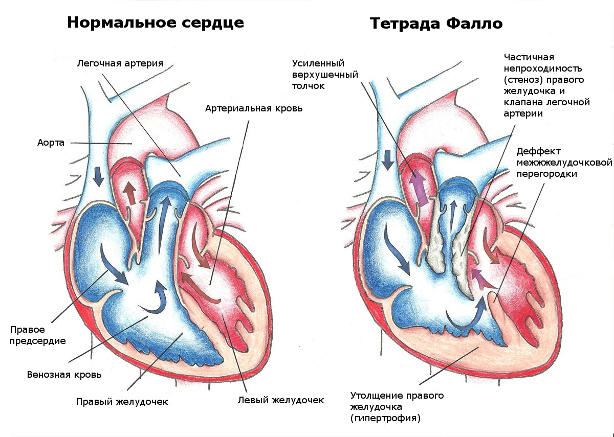 Tetralogy of Fallot
 Open heart surgery has two advantages.
- The surgeon has full access to the patient's heart.
- Such a surgical intervention is possible without state-of-the-art medical equipment.
However, there are also significant drawbacks.
- Surgical manipulations with the heart last several hours, which leads to fatigue of the operating team, during the operation there is a higher probability of making an erroneous action.
- Opening the chest is fraught with various injuries.
- There is a noticeable scar after heart surgery.
- Various complications are not excluded:
- myocardial infarction,
- thromboembolism,
- bleeding,
- infections;
- coma after surgery.
- A long recovery is required with significant limitations in the patient's activities.
In most cases, when surgery is performed with an opening of the chest, disability is given after heart surgery, as after a heart attack.
What operations and under what pathologies are performed on the open heart?
Pathologies of the coronary arteries
Coronary artery bypass grafting is done in case of serious atherosclerotic lesions of the coronary arteries, which led to a severe form of coronary heart disease. The essence of shunting is to create a bypass for blood flow to the heart using a shunt, for which an artery or vein taken from the patient is used. For example: mammary coronary artery bypass grafting (MCB) is performed using the internal mammary (mammary) artery.
 Operation Ross
 Heart valve defects
Today, valves made from the patient's biological material are used to replace damaged valves.
- The Ross procedure involves using the patient's own valvular pulmonary artery to replace a diseased aortic valve. An implant is placed in place of the pulmonary valve. Eliminates complications associated with rejection of a valve made of foreign material. Made for both adults and children.
- The Ozaki operation involves the use of the patient's own tissue. Only in this case, the replacement of the aortic valve is performed with a valve made from the patient's pericardium. Complications with valve rejection are not observed for the same reason.
Tuesday is surgery day. The team is preparing for a long morning work. During the operation, the chest is opened and the heart is prepared for vessel transplantation.
Disease history
Mr. Thomas, a 59-year-old tanker driver, is married with two adult children. He had shingles on the right side of his neck, followed by an uncomfortable constriction in his throat, accompanied by sweating and nausea. He first felt these symptoms while walking up the steps of his truck. They continued, and Thomas decided to seek the advice of a therapist.Thomas's high blood pressure, obesity, and long history of smoking were reason enough for an ECG. Her results showed the presence of coronary heart disease. Thomas was referred to a cardiac expert (a cardiac internist, not a surgeon). Despite the applied medical treatment, the pain continued.
Tests confirmed the presence of the disease, in particular an angiogram (a test using a dye injected into the artery to detect narrowing) revealed a narrowing in the left main coronary artery with damage to the left and right vessels. Since medical treatment was unsuccessful and angioplasty (stretching a narrowed vessel using a catheter) was not an option, Mr. Thomas was referred for surgery.
Monday
Mr. Thomas is hospitalized. His anamnesis, data of examinations and tests were analyzed. Two units of blood for transfusion are tested for compatibility. The patient is explained the essence of the operation and warned about the risk associated with it. Obtain written consent for CABG.Tuesday
Early in the morning, Mr. Thomas is being prepared for the operation.7:05 Premedication and anesthesia
8:15 a.m. Mr. Thomas was sedated 70 minutes ago and a ventilation tube has already been placed in his airway. After the application of anesthesia and paralyzing agents, his breathing is supported by a ventilator. Prior to transferring Mr. Thomas to the operating room, the anesthesiologist establishes monitoring of venous and arterial blood flow.8:16 OR Mr. Thomas is set up. On the left - a table with instruments, on the right - a ready-to-use heart-lung apparatus.
8:25 Patient in the operating room. The skin of his chest and legs are treated with an antiseptic solution to reduce the risk of infection.
8:40 Opening of the chest
The skin has already been processed, the patient is dressed in sterile clothes. One of the surgeons makes an incision in the leg to extract the vein, and the second cuts the skin on the chest. After a preliminary incision with an ordinary scalpel, he uses an electric one, which cuts the vessels, stopping the bleeding.8:48 The surgeon cuts the sternum bone with an electric saw with a pneumatic drive.
8:55 Artery and vein removal
View of the internal thoracic (mammary) artery in the mirror in the center of the surgical lamp. This artery is very elastic. The top end of it will remain in place, it will be cut off at the bottom and then connected to the coronary artery.An angled retractor is placed along the left edge of the sternum to lift it and expose the mammary artery that runs along the inside of the chest.
At the same time, one of the main veins on the leg - the great saphenous vein - is prepared for transplantation. It is almost completely removed from the left thigh.
9:05 Connecting to the heart-lung machine
The heart-lung machine is not yet connected to the patient. One of the five rotating pumps circulates the blood, while the rest are used as side pumps to transport separated blood to prevent blood loss during surgery. The patient needs to enter heparin - a means to thin the blood and prevent the formation of clots during its passage through plastic tubes.Tubes to the heart-lung apparatus. On the left, with bright red blood, is the arterial return line, which carries blood back into the patient's aorta. On the right - two tubes that drain blood from the inferior and superior vena cava under the influence of gravity. The incision in the sternum is fixed with a spacer.
Part of the heart-lung apparatus is a membrane oxygenating device that maintains blood circulation in the patient's body. At the moment, the device is filled with blood, carbon dioxide is removed from it. The blood is re-oxygenated and returned to the patient's body.
An arterial return tube is inserted into the aorta (the main artery of the body) and two venous drains are inserted into the vena cava (the main vein of the body).
9:25 Cardiac arrest
On the main artery - the aorta - a clamp is placed to isolate the heart from artificial blood circulation. Chilled fluid is injected into the isolated aorta to stop the heart. The surgeon puts on special glasses for microsurgery with loupes that give a magnification of 2.5 times. The blood vessels he will transplant are 2-3 mm in diameter, and the sutures are the diameter of a human hair.A thorough examination of the heart is carried out to confirm the data obtained using the angiogram. It is specified which coronary arteries need to be bypassed. It was decided to make two shunts.
After stopping the blood flow in the left anterior descending artery, a 1 cm long incision is made at the bypass site using a surgical loop.
10:00 First bypass
Close-up of the heart. The left internal mammary (mammary) artery - in the upper left corner - is sutured to the left anterior descending artery so that blood flow to the heart is restored. Arteries are hidden by epicardial fat.The end of the left internal mammary artery is sutured laterally to the left anterior descending artery. This forms the first bypass shunt.
The position of the first performed shunt. The end of the lower part of the left internal mammary artery - a blood vessel with a diameter of 3 mm - is completely sutured to the left anterior descending artery.
10:22 Second bypass
The second bypass shunt is sutured with the upper end to the aorta, and with the lower end to the right posterior descending artery. The transverse clamp is removed, blood flow through the heart is restored.The upper end of the venous shunt is connected to the aorta. Part of the aorta is isolated with an arcuate clamp and a hole is made into which a vein is sutured.
End of both bypass processes. The second shunt, shown on the left side of the diagram, is formed from the saphenous vein of the leg.
11:18 Chest closure
Circulation is restored, the heart contracts after an electric shock with the transition from ventricular fibrillation to sinus mode. Two drains are installed in the anterior and posterior parts of the heart. The blood thinning effect of heparin was eliminated by the drug protamine. The surgeon sews the separated halves of the sternum together. The skin will be closed with an internal absorbable suture.The nurse applies tape to the suture and to the drainage tubes leading from the patient's chest. Soon the patient will be placed in the intensive care unit, where he will be observed.
The human body. Outside and inside. №1 2008