The olfactory organ features physiological regeneration. Lecture on histology of sensory organs. Learning and teaching methods
The sense organs are the peripheral ends of the analyzers. The analyzer is an afferent link of the reflex arc, including a sensitive neuron of the sensory organ, associative afferent neurons and an associative efferent neuron of the cerebral cortex.
THE ANALYZER CONSISTS of 1) an end section where sensitive cells are located; 2) the intermediate part (represented by neurons along which the impulse moves to the center); 3) the central part is the cerebral cortex, in which the analysis and synthesis of the received information takes place and a response is prepared.
CLASSIFICATION OF SENSORY ORGANS. Sense organs are classified into 3 types: 1) Type I - eye and olfactory organ; 2) Type II - organs of hearing and taste and 3) Type III - receptors scattered throughout the body.
TYPE I ORGANS are characterized by the fact that they contain primary sensory neurons that develop from the brain vesicles, therefore they (these neurons) are called neurosensory.
TYPE II ORGANS are characterized by the fact that irritation is perceived not by neurons, but by sensitive epithelial cells developing from the skin ectoderm, which is why they are called sensoroepithelial. Sensitive epithelial cells transmit irritation to nerve cells, which are called secondary sensory cells. Sensitive epithelial cells have special hairs or microvilli.
VISUAL ORGAN
ORGAN OF VISUAL (oculus) is represented by the eyeball located in the orbit and an auxiliary apparatus. The auxiliary apparatus includes: eyelids, lacrimal apparatus and oculomotor muscles.
THE EYEBALL (bulbus oculi) contains three membranes. On the outside there is a fibrous membrane (tunica fibrosa), consisting of 2 parts: the anterior part (cornea) and the tunica albuginea, or sclera. Under the tunica albuginea is the choroid, and below it is the retina. The eyeball includes 3 systems (apparatus): 1) a dioptric or light-refracting apparatus, consisting of the cornea of the eye, the fluid of the anterior and posterior chambers of the eye, the lens and the vitreous body; 2) the accommodative apparatus, represented by the ciliary body and the ciliary girdle; this apparatus includes the iris, which should be classified as an adaptive apparatus; 3) light-perceiving apparatus, represented by the retina.
EYE DEVELOPMENT. The eye develops from several sources. 2 protrusions (eye vesicles) are formed from the brain vesicle. The anterior wall of the optic vesicles invaginates, as a result of which an optic cup is formed from each optic vesicle, connected to the neural tube by a hollow stalk and consisting of 2 walls: outer and inner. The pigment layer of the retina develops from the outer wall, and the neural layer of the retina develops from the inner wall. From the edges of the optic cup, the muscle constricting the pupil and the muscle dilating the pupil develop. The tunica albuginea, choroid, iris, ciliary body and connective tissue base of the cornea of the eye develop from mesenchyme. The anterior epithelium of the cornea and the lens develop from the cutaneous ectoderm. The development of the lens occurs as follows. At the time when the optic cup is formed, the cutaneous ectoderm located opposite the cup thickens and invaginates into the cup. This invagination separates from the ectoderm and during development turns into the lens. The vitreous body develops due to mesenchyme with the participation of blood vessels.
The fibrous membrane consists of a posterior part - the tunica albuginea and anterior part - the cornea. The tunica albuginea has a thickness of about 0.6 mm and consists of connective tissue plates, each of which is formed by a layer of parallel fibers. Between the plates there is the main intercellular substance, fibroblasts. At the border of the sclera and cornea there is Schlemm's canal (venous sinus), in which fluid circulates. Schlemm's canal drains fluid from the anterior chamber of the eye. FUNCTIONS of the sclera: 1) protective, 2) formative and 3) supporting, since the oculomotor muscles are attached to it.
DIOPTRIC EYE APPARATUS. The cornea has the shape of a convex-concave lens, i.e. collects rays, its refractive index is 1.37. The cornea consists of 5 layers: 1) anterior (outer) epithelium; 2) anterior limiting membrane (lamina limitans anterior); 3) proper substance of the cornea (substantia propria corneae); 4) posterior boundary layer (stratum limitans posterior); 5) posterior epithelium (epithelium posterioris).
The anterior epithelium is represented by a multilayered squamous non-keratinizing epithelium, including 3 layers: basal, spinous and flat. The epithelium is richly innervated by free nerve endings and is easily permeable to gases and liquid substances. The epithelium lies on a basement membrane, consisting of two layers: outer and inner.
THE ANTERIOR LIMITING PLATE (Bowman's membrane) is represented by an amorphous substance in which thin collagen fibrils pass. Its thickness is 6-10 microns.
THE PROPER SUBSTANCE of the cornea is represented by connective tissue plates consisting of parallel fibers. The plate consists of 1000 collagen fibers with a thickness of 0.3-0.6 microns. Between the plates there are fibroblasts and the main intercellular substance, rich in transparent sulfated glycosaminoglycans. The absence of blood vessels in the cornea and the presence of transparent sulfated glycosaminoglycans explain its transparency. The cornea is nourished by the blood vessels of the sclera and the fluid of the anterior chamber of the eye.
The POSTERIOR BORDER PLATE, about 10 µm thick, is represented by an amorphous substance in which a network of thin collagen fibrils is located.
POSTERIOR EPITHELIA is represented by one layer of flat polygonal epithelial cells.
The ANGLE OF THE ANTERIOR CHAMBER OF THE EYE is called differently: chamber, iridocorneal, because. located between the iris and cornea, and filtration, so fluid flows through it from the anterior chamber into Schlemm’s canal. In the sclera opposite the apex of the chamber angle there is a groove (sulcus scleralis internum). The posterior (outer) ridge of this groove is thickened. It is formed by circularly arranged collagen fibers. A ligamentous apparatus is attached to this place of the sclera, connecting the iris and ciliary body with the sclera. This device is also called trabecular. The trabecular apparatus has 2 parts: the corneoscleral (corneoscleral, or ligamentum corneascleralis) and the pectineal ligament (ligamentum pectinatum).
In the corneal-scleral part there are flattened trabeculae. In the center of each trabecula there is a collagen fiber, braided with elastic fibers and surrounded by a glassy mass. The trabeculae are covered with endothelium, which passes onto them from the posterior surface of the cornea. Between the trabeculae there are fountain spaces lined with endothelium. The fountain spaces carry out the outflow of fluid from the anterior chamber of the eye into Schlemm's canal.
Schlemm's canal is represented by a narrow fissure or several confluent fissures 2.5 mm wide and lined with endothelium. Anastomosing vessels extend from the outer edge of Schlemm's canal and drain into the veins of the sclera. This is the path of fluid outflow from the anterior chamber of the eye into the venous system.
The lens is located behind the anterior chamber of the eye in the center of the ciliary body ring and is fixed (attached) to the ciliary body using the ciliary girdle. The lens is located inside a thin transparent connective tissue capsule 11-18 microns thick. The collagen fibers of the ciliary girdle are attached to the edge of the capsule. The anterior surface of the lens is covered with single-layer squamous epithelium, which takes on a prismatic shape at its equator. The epithelium of the lens equator undergoes mitotic division (growth zone) and grows on its anterior and posterior surfaces. The epithelial cells of the posterior surface of the lens elongate as they mature and are called lens fibers (fibra lentis), consisting of a nucleus and cytoplasm. The cytoplasm contains the protein crystallin. The lens fibers are glued together using a substance that has the same refractive index as crystallin. The refractive index of the lens is 1.42.
During the process of differentiation, the lens fibers lose their nuclei and move to the center of the lens, forming its nucleus (nucleus lentis).
The lens has elasticity. It constantly strives to increase its curvature (round), but this is prevented by the collagen fibers of the ciliary girdle, which stretch the lens around its circumference.
The vitreous body (corpus vitreum) is located behind the lens and consists of the protein vitrein, located in the loops of a network of thin collagen fibers. In the central part vitreous less dense, the optic canal passes here, which approaches the macula - the place of best vision on the retina. The refractive index of the vitreous body is 1.33.
THE FUNCTION OF THE DIOPTRIC APPARATUS is to refract the rays and direct them to the macula of the retina.
THE ACCOMMODATION APPARATUS is represented by the ciliary body and the ciliary girdle, and a type of accommodative apparatus, the adaptive apparatus, is represented by the iris.
The CILIARY BODY (corpus ciliare) has the shape of a ring. The edge of the ring when cut has a triangular shape. The base of the triangle faces ventrally, the apex faces dorsally. The ciliary body consists of a ring (orbiculus ciliaris), located on the outside, and a ciliary crown (corona ciliaris). The ciliary body is covered with epithelium extending from the retina. The epithelium of the ciliary body is represented by 2 layers: 1) the basal layer consists of cubic pigment epithelial cells; 2) integumentary - from pigment-free epithelial cells of a prismatic shape. The surface of the epithelium is covered with a ciliary membrane (lamina). FUNCTION OF THE CILIAR BODY EPITHELIUM - participation in the secretion of fluid from the anterior and posterior chambers of the eye.
Ciliary processes (processus ciliaris) extend from the CILIARY CROWN, the basis of which is connective tissue in which small blood vessels pass.
CILIARY MUSCLE makes up the bulk of the ciliary body. It consists of bundles of smooth myocytes oriented in three directions: sagittally in the outer layer, circularly and radially in the inner layer.
CILAR GAND (zonula ciliaris) consists of collagen fibers arranged radially. The outer ends of these fibers are attached to the processes of the ciliary crown, the inner ends to the lens capsule. Thus, with the help of the ciliary girdle, the lens is fixed in the center of the ciliary body, which has the shape of a ring.
THE FUNCTION OF THE ACCOMMODATING APPARATUS OF THE EYE is accommodation, i.e. adaptation or adaptation of the eye to distance.
ADAPTATION OF THE EYE TO NEAR DISTANCE. When the eye is placed at a close distance, the ciliary muscle contracts. At the same time, the diameter of the ciliary body decreases, the tension of the collagen fibers of the ciliary girdle is weakened, the lens is rounded, i.e. its curvature increases and the focal length decreases.
WHEN SET YOUR EYES AT A FAR DISTANCE, the opposite happens. The ciliary muscle relaxes, the diameter of the ciliary body increases, the tension of the fibers of the ciliary girdle increases, the capsule
The lens stretches around the circumference, the lens flattens, i.e. its curvature decreases and the focal length increases. Thus, if the eye is placed at a close distance (reading a book), rapid fatigue occurs, since at this time the ciliary muscle is in a contracted state.
The vascular membrane of the eye (tunica vasculosa bulbi) is located medially from the sclera. Due to this membrane, the ciliary body and the iris are formed. There are 4 layers in the choroid: 1) the outer layer, it is called supravascular, (stratum supravasculare), consists of loose connective tissue, rich in pigment cells; 2) the vascular layer (stratum vasculare) consists of a plexus small arteries and veins, between which there are layers of connective tissue with numerous pigment cells; 3) the choriocapillary layer (lamina choriocapillaris) is formed by capillaries extending from the vessels of the vascular layer. The capillaries have different diameters along their length, turning into sinusoids. Between the capillary loops there are layers of connective tissue, pigment cells, fibroblasts; 4) the basal complex (complexus basalis) consists of a superficial collagen layer with a zone of elastic fibers, a deep layer formed by collagen fibers, and a basal membrane to which the epithelial cells of the pigment layer of the retina are adjacent. The thickness of the basal complex is 4 µm.
The FUNCTION of the choroid is trophic.
ADAPTATION APPARATUS OF THE EYE, which is an integral part of the accommodative apparatus, is represented by the iris and the pigment layer of the retina.
The IRIS (iris) has the shape of a disk, in the center of which there is a hole (pupil). The iris is closely connected with the ciliary body. There are 5 layers in the iris: 1) anterior (outer) epithelium (epithelium anterius iridis); 2) anterior boundary layer (stratum externum limitans); 3) vascular layer (stratum vasculosum); 4) internal boundary layer (stratum internum limitans); 5) posterior (inner) pigment layer (epithelium posterius pigmentosum).
The OUTER epithelium is represented by flattened, polygonal cells. They moved to the iris from the inner surface of the cornea.
The anterior (outer) boundary layer is characterized by the fact that it contains loose connective tissue rich in pigment cells. Depending on the quantity and quality of the pigment of the pigment cells, the eye has a certain color. If there is no pigment, then the iris will have a red color, since the blood vessels of the vascular layer will be visible through it.
The VASCULAR layer contains a plexus of small arteries and veins, between which the layers of connective tissue contain pigment cells.
The REAR boundary layer has the same structure as the front one. In the internal boundary layer there are 2 muscles: the muscle constrictor pupillae (musculus sphincter pupillae), which is innervated by fibers coming from the ciliary nerve ganglion, and the muscle dilatator pupillae, to which nerve fibers from the superior cervical sympathetic ganglion approach.
POSTERIOR EPITHELIA consists of 2 layers: the basal layer, consisting of cuboidal pigment epithelial cells, and elyocytes. This epithelium passes to the iris from the epithelium of the ciliary body.
FUNCTION OF THE IRIS - participation in light and dark adaptation of the eye. In bright light the pupil constricts, in low light it dilates.
RETINA (retina) - the light-receiving apparatus is located inward from the choroid. The retina has a photosensitive part, located in the back of the eye, and a non-photosensitive part, located closer to the ciliary body.
The light-sensitive layer of the retina includes a layer of pigment epithelium and a neural layer, which includes another 9 layers + pigment layer = 10 layers. The neural layer consists of a chain of three neurons: 1) photoreceptor (rod and cone), rod - cellula neurosensorius bacillifer, cone - cellula neurosensorius conifer; 2) associative neurons (bipolar, horizontal, amocrine); 3) ganglion or multipolar cells (neuronum multipolare). Due to the nuclear-containing parts of these neurons, 3 layers are formed, in particular, the bodies of light-sensitive neurons form the outer nuclear layer (stratum nuclearis externum); bodies of associative neurons - inner nuclear layer (stratum nuclearis internum); the bodies of ganglion neurons are the ganglion layer (stratum ganglionare). Due to the processes of these 3 neurons, 4 more layers are formed, in particular, the rods and cones of the dendrites of photoreceptor neurons form a layer of rods and cones (stratum photosensorium); axons of photoreceptor neurons and dendrites of associative neurons in places and synaptic connections together form the outer mesh layer (stratum plexiforme externum); axons of associative neurons and dendrites of ganlionic neurons in places of their synaptic connection form the internal mesh layer (stratum plexiforme internum); The axons of ganglion neurons form a layer of nerve fibers (stratum neurofibrarum).
Thus, 3 layers are formed due to the neuron bodies and 4 layers are formed due to the processes, for a total of 7 layers. Where are the other 3 layers? The eighth layer can be considered a layer of pigment cells (stratum pigmentosum). Where are the other 2 layers? The neuronal layer of the retina includes neuroglial cells, predominantly fibrous. They have an elongated shape and are located radially, therefore they are called radial (gliocytus radialis). The peripheral processes of radial gliocytes form a plexus between the layer of rod-cones and the outer nuclear layer. This plexus is called the outer glial limiting membrane (stratum limitans externum). The internal processes of these gliocytes, with their plexus, form the internal boundary layer (stratum limitans internum), located on the border with the vitreous body. Thus, due to the bodies of neurons, their processes, the pigment layer and processes of radial gliocytes, 10 layers are formed: 1) pigment layer; 2) layer of rods and cones; 3) outer boundary layer; 4) outer nuclear layer; 5) outer mesh layer; 6) inner nuclear layer; 7) inner mesh layer; 8) ganglion layer; 9) layer of nerve fibers; 10) internal boundary layer.
The human eye is called INVERTIVE. This means that the receptors of photoreceptor neurons (rods and cones) are directed not towards the light rays, but in the opposite direction. In this case, the rods and cones are directed towards the pigment layer of the retina. In order for a ray of light to reach the rods and cones, it needs to pass through the internal limiting layer, the nerve fiber layer, the ganglion layer, the internal reticularis layer, the internal nuclear layer, the outer reticularis layer, the outer nuclear layer, the outer limiting layer, and finally the rod and cone layer.
The location of the best vision of the retina is the macula flava. In the center of the spot there is a central fovea (fovea centralis). In the central fovea, all layers of the retina are sharply thinned, except for the outer nuclear layer, which consists mainly of the bodies of cone photoreceptor neurons, which are receptor devices for color vision. Inward from the macula there is a blind spot (macula cecum) - the papilla of the optic nerve (papilla nervi optici). The optic nerve papilla is formed by the axons of ganglion neurons included in the layer of nerve fibers. Thus, the axons of ganglion neurons form the optic nerve (nervus opticus).
STRUCTURE OF PHOTOSENSORY NEURONS (primary sensory cells).
ROD PHOTOSENSORIOUS NEURONS (neurocytus photosensorius bacillifer). Their bodies are located in the outer nuclear layer. The area of the body around the neuron nucleus is called the perikaryon. A central process, the axon, departs from the perikaryon, which ends in a synapse with the dendrites of associative neurons. The peripheral process, the dendrite, ends with a photoreceptor, the rod.
THE ROD OF A PHOTORECEPTOR NEURON consists of two segments, or segments: external and internal. The outer segment consists of disks, the number of which reaches 1000. Each disk is a double membrane. The thickness of the disk is 15 nm, the diameter is 2 mm, the distance between the disks is 15 nm, the distance between the membranes inside the disk is 1 nm. These disks are formed as follows. The cytolemma of the external segment is invaginated inward. A double membrane is formed. This double membrane is then laced off to form a disc. The disc membranes contain visual purple rhodopsin, which consists of the opsin protein and the vitamin A aldehyde retinal. Thus, vitamin A is required for the rods to function.
The outer segment is connected to the inner segment by means of a cilium, consisting of 9 pairs of peripheral microtubules and one pair of central microtubules. Microtubules are attached to the basal body. The INNER MEMBER contains general organelles and enzymes. Rods perceive black and white color and are twilight vision devices. The number of rod neurons in the human retina is about 130 million. The length of the largest rods reaches 75 microns.
CONE PHOTORECEPTOR NEURONS consist of a perikaryon, an axon (central process) and a dendrite (peripheral process). The axon enters into synaptic communication with associative neurons of the retina, the dendrite ends in a photoreceptor called a cone. CONES differ from rods in the structure, shape and content of visual purple, which in cones is called iodopsin.
The outer member of the cone consists of 1000 half-discs. Half-discs are formed by invagination of the cytolemma of the outer segment and are not detached from it. Therefore, the hemidiscs remain connected to the cytolemma of the outer segment. The outer segment is connected to the inner segment using a cilium.
THE INNER MEMBER OF THE CONE includes general organelles, enzymes and an ellipsoid consisting of a lipid droplet surrounded by a dense layer of mitochondria. Ellipsoids play a role in color perception. The number of cone photoreceptor neurons in the human retina is about 6-7 million; they are color vision devices. Depending on what type of pigment is contained in the membranes of the cones, some of them perceive red, others blue, and others green. With the combination of these three types of cones, the human eye is able to perceive all the colors of the rainbow. The presence or absence of a particular pigment in cones depends on the presence or absence of the corresponding gene on the sex X chromosome.
If there is no pigment that perceives red color, this is called protanopia, and green color is called deuteranopia.
ASSOCIATIVE NEURONS OF THE RETINA (bipolar, horizontal and amocrine)
BODIES OF BIPOLAR NEUROCYTES (neurocytus bipolaris) are located in the inner nuclear layer. Their dendrites contact the axons of several rod neurons and one cone neuron, and the axons contact the dendrites of ganglion neurons. Thus, bipolar neurons transmit visual impulses from photoreceptor neurons to ganglion neurons.
THE BODIES OF HORIZONTAL NEURONS are located in the inner nuclear layer closer to the photoreceptor neurons. The dendrites of horizontal neurons contact the axons of photoreceptor neurons, their long axons go in the horizontal direction and form axo-axonal (inhibitory) synapses with several photoreceptor cells. Thanks to horizontal neurons, the impulse coming in the central part is transmitted to the bipolar cells, and the impulse passing laterally from the center is inhibited in the area of axo-axonal synapses. This is called lateral inhibition, which ensures the clarity and contrast of the image on the retina.
The bodies of amocrine neurons are located in the inner nuclear layer closer to the ganglion cells. Amocrine cells contact ganglion neurons and perform the same function as horizontal neurons, but only in relation to ganglion neurons.
GANGLIONAR (MULTIPOLAR) NEUROCYTES are located in the ganglion layer of the retina. Their dendrites contact the axons of bipolar neurocytes and amocrine cells, and the axons form a layer of nerve fibers that join together in the area of the optic nerve to form the optic nerve.
THE VISUAL PATHWAY starts from the receptors of photoreceptor neurons (rods and cones), where, under the influence of light rays, a chemical reaction begins with the subsequent disintegration of the visual pigment, an increase in the permeability of the cytolemma of rods and cones occurs, which results in a light impulse. This impulse is transmitted to the bipolar neuron, then to the ganglion neuron, and then to its axon. The optic nerve is formed from the axons of ganglion neurons, along which the impulse is directed towards the central nervous system. Through the optic foramen (foramen opticum), the optic nerve enters the cranial cavity and approaches the optic chiasm (hiasma opticum). Here the inner halves of the nerve cross, the outer halves go without crossing. The visual tract (tractus opticus) begins from the optic chiasm. Included optic tract the axons of the ganglion neurons of the retina are directed to the 4th neuron, located in the pads of the visual tuberosities, the lateral geniculate bodies and in the superior colliculi of the quadrigemina, the axons of the fourth neurons, located in the cushions of the optic tuberosities and the latral geniculate bodies, are sent to the calcarine sulcus of the cerebral cortex, where the central end of the visual analyzer.
THE PIGMENT LAYER OF THE RETINA consists of 6 million pigment cells, which with their basal surface lie on the basement membrane of the choroid. The light cytoplasm of pigment cells (melanocytes) is poor in organelles of general importance and contains a large amount of pigment (melanosomes). Melanocyte nuclei are spherical. Processes (microvilli) extend from the apical surface of melanocytes, which extend between the ends of the rods and cones. Each rod is surrounded by 6-7 such processes, each cone is surrounded by 40 processes. The pigment of these cells is able to migrate from the cell body to the processes and from the processes to the body of the melanocyte. This migration occurs under the influence of the melanocyte-stimulating hormone of the intermediate part of the adenohypophysis and with the participation of filaments inside the cell itself.
THE FUNCTIONS OF THE RETINA PIGMENT LAYER are numerous. 1. It is an integral part of the adaptive apparatus of the eye. 2. Participates in the inhibition of peroxide oxidation. 3Performs a phagocytic function.4.Participates in the metabolism of vitamin A.
PARTICIPATION OF THE PIGMENT LAYER IN EYE ADAPTATION. In bright light, too much light rays reach the cones and rods of the retina. In this case, the pupil instantly narrows to reduce the number of rays. But the eye feels uncomfortable. Then the pigment from the cell bodies begins to migrate into the processes located between the rods and cones. As a result of this, a so-called pigmented beard is formed. Since the rods do not participate in the perception of color vision, they become longer and sink even deeper into the pigment beard. At this time, the cones shorten so that the rays fall on them. Thus, the pigment beard, like a screen, covers the sticks from light rays. At this time, the eye does not experience any unpleasant sensations.
In LOW LIGHT, the pupil immediately dilates, but the eye does not see objects well. After some time, the contours of objects appear more clearly. During this time, the following changes occurred in the pigment layer of the retina. The pigment from the processes returns back to the pigment cell bodies, i.e. The pigmented beard decreases or completely disappears. Since the cones are not involved in the perception of black and white color, they elongate and are immersed in a short pigment beard. On the contrary, the rods shorten somewhat and recede from the pigment layer so that greatest number rays in low light fell on the outer segment of the rods. At this moment, a person begins to clearly see objects in a poorly lit room.
PARTICIPATION OF THE PIGMENT LAYER IN THE INHIBITION OF PEROXIDATION is carried out in 2 ways: 1) due to the fact that the enzymes catalase and peroxidase are released from the peroxisomes of pigment cells, which inhibit peroxide oxidation; 2) on the surface of the pigment granules, adsorption of metal molecules involved in catalyzing peroxide oxidation occurs.
PARTICIPATION OF THE PIGMENT LAYER IN THE METABOLISM OF VITAMIN A (retinol). Retinol is deposited in the liver. To deliver retinol to the retina, retinol-binding protein is synthesized in the liver. Vitamin A, or retinol, enters the bloodstream and is transported through the bloodstream to the pigment layer of the retina. Vitamin A molecules are captured by pigment cell receptors and penetrate into the cell, in which rhodopsin is synthesized, which then enters the membranes of the discs of the outer segments of the rods.
PHAGOCYTIC FUNCTION OF THE PIGMENT LAYER. Pygmeniocytes phagocytose rod discs and cone half-discs. During the day, approximately 80 disks of each rod and 80 half-discs of the cone are phagocytosed.
REGENERATION OF CONES AND RODS is carried out as follows. First, aging occurs in the apical discs of rods and half-discs of cones. At the base of the outer segments of rods and cones, their cytolemma grows, which then invaginates into the segment, resulting in the formation of about 80 new discs and half-discs in each outer segment. Old degenerative discs and hemidiscs are phagocytosed by pigment cells. Thus, in the outer segment of each rod or cone, about 80 new discs and half-discs are formed every day and the same number are phagocytosed by pigmentocytes. As a result of this, the rod disks or cone half-discs are renewed within approximately 12 days.
The process of formation of new discs and half-discs and their phagocytosis is carried out in accordance with daily, or circadian rhythms: rod discs are destroyed and phagocytosed during the daytime (when they do not function); cones, on the contrary, at night, when their function ceases. It depends on some factors. In particular, during the daytime, when the rods do not function, a large amount of vitamin A accumulates in their discs, which promotes the destruction of the discs (has membranolytic properties). The second factor is cAMP (cyclic adenosine monophosphate). It inhibits the destruction of discs, but in the daytime there is little cAMP, so the process of their destruction and phagocytosis is not suppressed. In the dark, the amount of cAMP increases, therefore, the inhibition of the destruction and phagocytosis of rods increases, i.e. the destruction of rod discs at night is weakened or stops completely.
AUXILIARY APPARATUS OF THE EYE is represented by eyelids, lacrimal apparatus and extraocular muscles.
The eyelids are covered on the outside with skin (skin surface), on the inside - by the conjunctiva, which is lined with stratified squamous epithelium and continues into the conjunctiva of the eye. In the thickness of the eyelid, closer to the posterior surface, there is a torsal plate consisting of dense connective tissue. Closer to the anterior surface lies the annular muscle. The tendons of the levator palpebral muscle are also located here.
Along the edge of the eyelid there are 2-3 rows of eyelashes. Several excretory ducts of the sebaceous glands open into the funnel of the eyelash root. The ducts of modified sweat glands (ciliary glands) also open here. In the thickness of the torsal plate there are sebaceous glands (meibomian glands), the excretory ducts of which open along the edge of the eyelid. In the inner corner of the eye there is a rudimentary eyelid, covered with stratified squamous epithelium, which contains mucous cells.
The lacrimal apparatus of the eye consists of the lacrimal glands, the lacrimal sac and the nasolacrimal canal. LACRIMAL GLANDS are represented by several complex branched alveolar-tubular glands; they produce a secretion consisting of water, chlorides (1.5%), albumin (0.5%) and mucus. Tear fluid contains lysozyme, which destroys bacteria.
The lacrimal sac and the nasolacrimal canal are lined with double or multi-row epithelium. The ducts of the lacrimal glands flow into the lacrimal sac.
THE OLfactory ORGAN is represented by olfactory fields located in the superior and partially middle turbinate. The olfactory organ DEVELOPES in early embryogenesis from olfactory placodes (thickenings of the ectoderm near the head end of the neural tube). Olfactory pits are formed from the placodes, which migrate to the area of the superior and middle nasal concha. Here, as a result of differentiation of the olfactory pits, olfactory and supporting cells are formed. During the differentiation of olfactory cells, they form a dendrite and an axon. The axons of the olfactory cells travel to the brain.
OLfactory fields are presented in the form of multirow olfactory epithelium lying on a rather thick basement membrane. Among the olfactory cells there are: 1) olfactory cells (epitheliocytus olfactorius); 2) supporting cells (epitheliocytus sustentans) and 3) basal cells (epitheliocytus basalis).
OLfactory cells are neurons that have a dendrite and an axon. DENDRITE is directed to the periphery, i.e. on the surface of the olfactory spot and ends with a thickening - a club (clava olfactoria). The club is covered with motile cilia, on the cytolemma of which there are receptor proteins that perceive odors. Receptor proteins capture molecules of odorous substances, which dissolve and a chemical reaction begins, causing a change in the permeability of the cytolemma and the occurrence of an impulse.
The axon of the olfactory cell through the ethmoid bone is sent as part of bundles (fila olfactorica) to the olfactory bulb (bulbus olfactorius) - the subcortical olfactory center of the brain stem, where the mitral neurons are located. The axons of mitral neurons are sent to the ancient cortex (hippocampus) and to the hypocampal gyrus of the neocortex (new cortex), where the cortical olfactory center is located. In the middle part of the olfactory cells there is a nucleus; the neuroplasm contains mitochondria, the Golgi complex, and granular ER.
SUPPORTING CELLS have a prismatic shape, their basal end lies on the basement membrane, the apical end extends to the surface of the olfactory field, the nucleus is located in the center of the cell. Organelles of general importance are well developed, there are microfilaments and secretory granules. FUNCTION - they secrete a liquid secretion of the apocrine type, in which odorous substances dissolve, and isolate the olfactory cells from each other.
BASAL CELLS are triangular in shape, poorly differentiated in function, due to them the olfactory cells are renewed every 30 days.
OLfactory GLANDS are located under the basement membrane in loose connective tissue, have a tubular structure, and produce a liquid secretion that dissolves odorous substances.
The VOMERONASAL ORGAN is located in the form of two tubes in the lower part of the nasal septum.
DEVELOPMENT. At the 6th week of embryogenesis, the epithelium of the base of the nasal septum in the form of two tubes grows into the connective tissue. At the 7th week, a round cavity of the tubules of the vomeronasal organ is formed. At week 21, its sensory and supporting cells differentiate. A peripheral process departs from the body of the sensory cells, the end of which thickens in the form of a club; the second process, the axon, unites with the same processes, resulting in the formation of bundles that enter the brain through the cribriform plate.
STRUCTURE OF THE VOMERONASAL ORGAN. The anterior (distal) end of the tubules of the vomeronasal organ ends blindly, while the posterior (proximal) end opens into the nasal cavity. The epithelium of the vomeronasal organ is represented by three types of cells: 1) sensory, 2) sustentocytes and 3) basal.
SENSOEPITHELIAL CELLS have an elongated shape, contain an oval nucleus and organelles of general importance. A peripheral process extends from their body, ending in a thickening, or club (clava olfactoria). Motionless microvilli extend from the club, into the cytolemma of which receptor proteins are built in, perceiving the odor secreted by the glands of the reproductive system of the opposite individual. The central process of sensory cells unites with other similar processes into unmyelinated cable-type fibers and through the cribriform plate is directed to the brain and carries a nerve impulse to the accessory olfactory bulb.
SUSTENTOCYTES of the vomeronasal organ have an elongated shape, an oval nucleus. Their cytoplasm contains the Golgi complex, EPS, and mitochondria. There are microvilli on the apical surface. These cells secrete a liquid secretion that dissolves odorant molecules.
BASAL CELLS are poorly differentiated. Due to the differentiation of these cells, the renewal of sensoroepithelial cells and sustentocytes occurs.
THE FUNCTIONAL IMPORTANCE of the vomeronasal organ lies in its influence on sexual behavior and the emotional state of a person.
Chapter 12. SENSE ORGANS
Chapter 12. SENSE ORGANS
12.1. GENERAL MORPHOFUNCTIONAL CHARACTERISTICS AND CLASSIFICATION
The sense organs provide the perception of various stimuli acting on the body; transformation and encoding of external energy into a nerve impulse, transmission along nerve pathways to the subcortical and cortical centers, where the analysis of received information and the formation of subjective sensations occur. Sense organs are analyzers of the external and internal environment that ensure the body’s adaptation to specific conditions.
Accordingly, each analyzer has three parts: peripheral (receptor), intermediate And central.
Peripheral part represented by organs in which specialized receptor cells are located. According to the specificity of perception of stimuli, there are mechanoreceptors (receptors of the organ of hearing, balance, tactile receptors of the skin, receptors of the movement apparatus, baroreceptors), chemoreceptors (organs of taste, smell, vascular interoreceptors), photoreceptors (retina), thermoreceptors (skin, internal organs), pain receptors.
Intermediate (conductor) part The analyzer is a chain of interneurons through which the nerve impulse from the receptor cells is transmitted to the cortical centers. On this path there may be intermediate, subcortical centers where afferent information is processed and switched to efferent centers.
central part analyzer is represented by areas of the cerebral cortex. The center analyzes the received information and forms subjective feelings. Here information can be stored in long-term memory or switched to efferent pathways.
Classification of sense organs. Depending on the structure and function of the receptor part, sensory organs are divided into three types.
To the first type These include the sense organs, whose receptors are specialized neurosensory cells (the organ of vision, the organ of smell), which convert external energy into a nerve impulse.
To the second type These include sensory organs whose receptors are not nerve cells, but epithelial cells (sensoepithelial). From them
the converted irritation is transmitted to the dendrites of sensory neurons, which perceive the excitation of the sensoroepithelial cells and generate a nerve impulse (organs of hearing, balance, taste).
To the third type include the proprioceptive (musculoskeletal) cutaneous and visceral sensory systems. The peripheral sections in them are represented by various encapsulated and non-encapsulated receptors (see Chapter 10).
12.2. VISUAL ORGAN
Eye (ophthalmos oculus)- the organ of vision, which is the peripheral part of the visual analyzer, in which the receptor function is performed by the neurosensory cells of the retina.
12.2.1. Eye development
The eye develops from various embryonic rudiments (Fig. 12.1). The retina and optic nerve are formed from the neural tube by first forming the so-called eye vesicles, maintaining connection with the embryonic brain using hollow eyestalks. The anterior part of the optic vesicle protrudes into its cavity, due to which it takes the shape of a double-walled optic cup. The part of the ectoderm located opposite the opening of the optic cup thickens, invaginates and laces off, giving rise to the primordium lens The ectoderm undergoes these changes under the influence of differentiation inducers formed in the optic vesicle. Initially, the lens has the appearance of a hollow epithelial vesicle. Then the epithelial cells of its posterior wall elongate and turn into so-called lens fibers, filling the cavity of the bubble. During development, the inner wall of the optic cup is transformed into retina, and the outer one - in pigment layer retina. At the 4th week of embryogenesis, the retinal rudiment consists of homogeneous poorly differentiated cells. At the 5th week, a division of the retina into two layers appears: the outer (from the center of the eye) - nuclear, and the inner layer, which does not contain nuclei. The outer nuclear layer plays the role of a matrix zone, where numerous mitotic figures are observed. As a result of subsequent divergent differentiation of stem (matrix) cells, cellular differentiates of various layers of the retina develop. Thus, at the beginning of the 6th week, neuroblasts begin to move out of the matrix zone, forming the inner layer. At the end of the 3rd month, a layer of large ganglion neurons. Last of all, the outer nuclear layer appears in the retina, consisting of neurosensory cells - rods And cone neurons. This happens shortly before birth. In addition to neuroblasts, the matrix layer of the retina produces glioblasts- sources of development of glial cells.
Rice. 12.1. Eye Development:
a-c - sagittal sections of the eyes of embryos on various stages development. 1 - ectoderm; 2 - lens placode - future lens; 3 - optic vesicle; 4 - vascular notch; 5 - outer wall of the optic cup - future pigment layer of the retina; 6 - inner wall of the optic cup; 7 - stalk - future optic nerve; 8 - lens vesicle
Among them become highly differentiated radial gliocytes(Müllerian fibers) penetrating the entire thickness of the retina.
The stalk of the optic cup is penetrated by axons formed in the retina ganglion multipolar neurons. These axons form the optic nerve, which goes to the brain. From the surrounding optic cup mesenchyme forms choroid And sclera. In the anterior part of the eye, the sclera becomes transparent, covered with stratified squamous epithelium (ectodermal). cornea. The inside of the cornea is lined with single-layer epithelium of neuroglial origin. Vessels and mesenchyme penetrating into early stages development inside the optic cup, together with the embryonic retina, take part in the formation vitreous And irises. The iris muscle that constricts the pupil develops from the marginal thickening of the outer and inner layers of the optic cup, and muscle that dilates the pupil- from the outer leaf. Thus, both muscles of the iris are neural in origin.
12.2.2. Structure of the eye
Eyeball (bulbus oculi) consists of three shells. Outer (fibrous) membrane eyeball (tunica fibrosa bulbi), to which the external muscles of the eye are attached, provides a protective function. It distinguishes the anterior transparent section - cornea and the posterior opaque section - sclera Middle (choroid) membrane (tunica vasculosa bulbi) plays a major role in metabolic processes. It has three parts: part of the iris, part of the ciliary body and the vascular part itself - the choroid (choroidea).
Inner lining of the eye- retina (tunica interna bulbi, retina)- sensory, receptor part of the visual analyzer in which

Rice. 12.2. Structure of the anterior section of the eyeball (diagram):
1 - cornea; 2 - anterior chamber of the eye; 3 - iris; 4 - posterior chamber of the eye; 5 - lens; 6 - ciliary girdle (ligament of Zinn); 7 - vitreous body; 8 - pectineal ligament; 9 - venous sinus of the sclera; 10 - ciliary (ciliary) body: A- processes of the ciliary body; b- ciliary muscle; 11 - sclera; 12 - choroid; 13 - jagged line; 14 - retina
under the influence of light, photochemical transformations of visual pigments, phototransduction, changes in the bioelectrical activity of neurons and transmission of information about outside world to the subcortical and cortical visual centers.
The membranes of the eye and their derivatives form three functional apparatuses: light refractive, or dioptric (cornea, fluid of the anterior and posterior chambers of the eye, lens and vitreous body); accommodative(iris, ciliary body with ciliary processes); receptor apparatus (retina).
The outer fibrous membrane is the sclera(sclera), formed by dense, shaped fibrous connective tissue containing bundles of collagen fibers, between which there are flattened fibroblasts and individual elastic fibers (Fig. 12.2). Bundles of collagen fibers, becoming thinner, pass into the cornea's own substance.
The thickness of the sclera in the posterior section around the optic nerve is greatest - 1.2-1.5 mm; anteriorly the sclera thins to 0.6 mm at the equator and to 0.3-0.4 mm behind the insertion of the rectus muscles. In the region of the optic nerve head, most (2/3) of the thinned fibrous membrane merges with the optic nerve sheath, and the thinned inner layers form the cribriform plate (lamina cribrosa). With an increase in intraocular pressure, the fibrous membrane becomes thinner, which is the cause of some pathological changes.
Light refractive apparatus of the eye
The refractive (dioptric) apparatus of the eye includes the cornea, lens, vitreous body, and fluid (aqueous humor) of the anterior and posterior chambers of the eye.
Cornea(cornea) occupies 1/16 of the area of the fibrous membrane of the eye and, performing a protective function, is characterized by high optical homogeneity, transmits and refracts light rays and is an integral part of the light-refracting apparatus of the eye.

Rice. 12.3. Cornea of the eye: 1 - stratified squamous non-keratinizing epithelium; 2 - anterior border plate; 3 - intrinsic substance; 4 - posterior border plate; 5 - posterior corneal epithelium
The thickness of the cornea is 0.8-0.9 microns in the center and 1.1 microns at the periphery, the radius of curvature is 7.8 microns, the refractive index is 1.37, the refractive power is 40 diopters.
Microscopically, five layers are distinguished in the cornea: 1) anterior multilayered squamous non-keratinizing epithelium; 2) anterior limiting plate (Bowman’s membrane); 3) own substance; 4) posterior limiting plate (Descemet's membrane); 5) posterior epithelium (endothelium of the anterior chamber) (Fig. 12.3).
Cells anterior corneal epithelium (keratocytes) tightly adjacent to each other, arranged in five layers, connected by desmosomes (see Fig. 12.3). The basal layer is located on the anterior limiting plate. Under pathological conditions (if the connection between the basal layer and the anterior limiting plate is not strong enough), detachment of the basal layer from the limiting plate occurs. The cells of the basal layer of the epithelium (cambial) have a prismatic shape and an oval nucleus located close to the top of the cell. Adjacent to the basal layer are 2-3 layers of multifaceted cells. Their laterally elongated processes are embedded between neighboring epithelial cells, like wings (winged, or spiny, cells). Roof nuclei
The patched cells are round. The two superficial epithelial layers consist of sharply flattened cells and do not show signs of keratinization. The elongated narrow nuclei of the cells of the outer layers of the epithelium are located parallel to the surface of the cornea. The epithelium contains numerous free nerve endings, which determine the high tactile sensitivity of the cornea. The surface of the cornea is moistened with the secretion of the lacrimal and conjunctival glands, which protects the eye from the harmful physical and chemical effects of the outside world and bacteria. The corneal epithelium has a high regenerative capacity. Under the corneal epithelium there is a structureless anterior border plate (lamina limitans anterior)- Bowman's membrane- thickness 6-9 microns. This is a homogeneous layer of randomly located collagen fibrils - a product of the vital activity of epithelial cells. The boundary between Bowman's membrane and the epithelium is well defined; the fusion of Bowman's membrane with the stroma occurs imperceptibly.
Proper substance of the cornea (substantia propria cornea)- stroma- consists of homogeneous thin connective tissue plates, intersecting at an angle, but regularly alternating and located parallel to the surface of the cornea. Processed flat cells, which are types of fibroblasts, are located in the plates and between them. The plates consist of parallel bundles of collagen fibrils with a diameter of 0.3-0.6 microns (1000 in each plate). Cells and fibrils are immersed in a ground substance rich in glycosaminoglycans (mainly keratin sulfates), which ensures the transparency of the cornea's own substance. The optimal concentration of water in the stroma (75-80%) is maintained by the mechanism of transport of sodium ions through the posterior epithelium. The transition of the transparent cornea to the opaque sclera occurs in the area limbo cornea (limbus corneae). The cornea itself does not have blood vessels.
Posterior border plate (lamina limitans posterior)- Descemet's membrane- 5-10 microns thick, represented by collagen fibers with a diameter of 10 nm, immersed in an amorphous substance. This is a glassy structure that strongly refracts light. It consists of two layers: the outer - elastic, the inner - cuticular and is a derivative of posterior epithelial cells. Characteristic Features The posterior border plate is strength, resistance to chemical agents and the melting effect of purulent exudate in corneal ulcers.
When the anterior layers die, Descemet's membrane protrudes into a transparent vesicle (descemetocele). At the periphery, it thickens, and in elderly people, round warty formations - Hassall-Henle bodies - can form in this place.
At the limbus, Descemet's membrane, thinning and becoming fibrous, passes into the trabecular apparatus of the sclera (see below).
Posterior epithelium (epithelium posterius), or endothelium of the anterior chamber, consists of a single layer of hexagonal cells. The cell nuclei are round or slightly oval, their axis is parallel to the surface of the cornea. Cells often contain vacuoles. At the periphery of the cornea, the posterior epithelium passes directly onto the fibers of the trabecular meshwork, forming the outer covering of each trabecular fiber, extending in length. The posterior epithelium protects the cornea from moisture from the anterior chamber.
Metabolic processes in the cornea are ensured by the diffusion of nutrients from the anterior chamber of the eye due to the marginal looped network of the cornea, numerous terminal capillary branches forming a dense perilimbal plexus.
The lymphatic system of the cornea is formed from narrow lymphatic slits communicating with the ciliary venous plexus.
The cornea is different high sensitivity, which is explained by the presence of nerve endings in it. Long ciliary nerves, representing branches of the nasociliary nerve arising from the first branch trigeminal nerve, on the periphery of the cornea penetrate into its thickness, lose myelin at some distance from the limbus, dividing dichotomously. The nerve branches form the following plexuses: in the substance of the cornea, preterminal and under the anterior border plate - terminal, subbasal (Riser's plexus).
During inflammatory processes, blood capillaries and cells (leukocytes, macrophages, etc.) penetrate from the limbus into the cornea's own substance, which leads to its clouding and keratinization, the formation of a cataract.
Front camera formed by the cornea (outer wall) and the iris (posterior wall), in the area of the pupil - by the anterior capsule of the lens. At its extreme periphery in the corner of the anterior chamber there is an iridocorneal (chamber) angle (spatia anguli iridocornealis) with a small area of the ciliary (ciliary) body. The chamber (so-called filtration) corner borders the drainage apparatus - Schlemm's canal. The state of the chamber angle plays a large role in the exchange of aqueous humor and in changes in intraocular pressure. Corresponding to the apex of the angle, a ring-shaped groove runs through the sclera (sulcus sclerae internus). The posterior edge of the groove is somewhat thickened and forms a scleral ridge formed by circular fibers of the sclera (posterior limiting ring of Schwalbe). The scleral ridge serves as the attachment point for the suspensory ligament of the ciliary body and the iris, a trabecular apparatus that fills the anterior part of the scleral groove. In the posterior part it covers Schlemm's canal.
Trabecular apparatus, formerly erroneously called the pectineal ligament, consists of two parts: sclerocorneal (lig. sclerocorneale), occupying most of the trabecular apparatus, and the second, more delicate - uveal part, which is located on the inside and is actually pectineal ligament (lig. pectinatum). The sclerocorneal section of the trabecular apparatus is attached to the scleral spur and partially merges with the ciliary muscle (Brücke muscle). The sclerocorneal part of the trabecular apparatus consists of a network of trabeculae with a complex structure.
In the center of each trabecula, which is a flat thin cord, there passes a collagen fiber, entwined, reinforced with elastic fibers and covered on the outside with a case of a homogeneous vitreous membrane, which is a continuation of the posterior border plate. Numerous free slit-like openings remain between the complex interweaving of corneoscleral fibers - fountain spaces, lined by the endothelium of the anterior chamber, passing from the posterior surface of the cornea. Fountain spaces are directed towards the wall venous sinus of the sclera (sinus venosus sclerae)- Schlemm's canal, located in the lower part of the scleral groove, 0.25 cm wide. In some places it is divided into a number of tubules, then merging into one trunk. The inside of Schlemm's canal is lined with endothelium. Wide, sometimes varicose vessels extend from its outer side, forming a complex network of anastomoses, from which veins originate, draining aqueous humor from the anterior and posterior chambers into the deep scleral venous plexus.
Lens(lens). This is a transparent biconvex body, the shape of which changes during the accommodation of the eye to see near and distant objects. Together with the cornea and vitreous body, the lens constitutes the main light-refracting medium. The radius of curvature of the lens varies from 6 to 10 mm, the refractive index is 1.42. The lens is covered with a transparent capsule 11-18 microns thick. This is the basement membrane of the epithelium, which contains collagen, sulfated glycosaminoglycan, etc. The anterior wall of the lens consists of single-layer squamous epithelium (epithelium lentis). Towards the equator, epithelial cells become taller and form germinal zone lens This zone is the cambial zone for the cells of the anterior and posterior surfaces of the lens. New epithelial cells transform into lens fibers (fibrae lentis). Each fiber is a transparent hexagonal prism. In the cytoplasm of the lens fibers there is a transparent protein - crystallin. The fibers are glued together with a special substance that has the same refractive index as them. The centrally located fibers lose their nuclei, shorten and, overlapping each other, form the nucleus of the lens.
The lens is supported in the eye by fibers ciliary girdle (zonula ciliaris), formed by radially arranged bundles of inextensible fibers attached on one side to the ciliary (ciliary) body, and on the other to the lens capsule, due to which the contraction of the muscles of the ciliary body is transmitted to the lens. Knowledge of the laws of the structure and histophysiology of the lens made it possible to develop methods for creating artificial lenses and widely introduce their transplantation into clinical practice, which made possible treatment patients with clouding of the lens (cataract).
Vitreous body(corpus vitreum). This is a transparent mass of jelly-like substance that fills the cavity between the lens and the retina, containing 99% water. On fixed preparations, the vitreous body has a mesh structure. At the periphery it is denser than in the center.
A canal passes through the vitreous body - a remnant of the embryonic vascular system of the eye - from the retinal papilla to the posterior surface of the lens. The vitreous body contains the protein vitrein and hyaluronic acid; the cells found in it are hyalocytes, macrophages and lymphocytes. The refractive index of the vitreous body is 1.33.
Accommodative apparatus of the eye
The accommodative apparatus of the eye (iris, ciliary body with ciliary belt) ensures a change in the shape and refractive power of the lens, focusing the image on the retina, as well as adapting the eye to the intensity of light.
Iris(iris). It is a disc-shaped formation with a hole of variable size (pupil) in the center. It is a derivative of the choroid (mainly) and retina. At the back, the iris is covered with retinal pigment epithelium. Located between the cornea and lens on the border between the anterior and posterior chambers of the eye (Fig. 12.4). The edge of the iris connecting it to the ciliary body is called the ciliary (ciliary) edge. The iris stroma consists of loose fibrous connective tissue rich in pigment cells. Myoneural cells are located here. The iris carries out its function as the diaphragm of the eye with the help of two muscles: the constrictor (musculus sphincter pupillae) and expanding (musculus dilatator pupillae) pupil.
There are five layers in the iris: anterior (outer) epithelium, covering the anterior surface of the iris, anterior border (outer avascular) layer, vascular layer, posterior (inner) border layer And posterior (pigment) epithelium.
Anterior epithelium (epithelium anterius iridis) represented by neuroglial flat polygonal cells. It is a continuation of the epithelium covering the posterior surface of the cornea.
Anterior boundary layer (stratum limitans anterius) consists of a ground substance in which a significant number of fibroblasts and pigment cells are located. The different positions and numbers of melanin-containing cells determine eye color. Albinos have no pigment and the iris is red due to the fact that blood vessels are visible through its thickness. In old age, depigmentation of the iris is observed, and it becomes lighter.
Vascular layer (stratum vasculosum) consists of numerous vessels, the space between which is filled with loose fibrous connective tissue with pigment cells.
Posterior boundary layer (stratum limitans posterius) does not differ in structure from the anterior layer.
Posterior pigment epithelium (epithelium posterius pigmentosum) is a continuation of the double-layer retinal epithelium covering the ciliary body and processes. It includes differons of modified gliocytes and pigmentocytes.
Ciliary, or ciliary, body(corpus ciliare). The ciliary body is a derivative of the choroid and retina. Performs the function of fixing the lens and changing its curvature, thereby participating in the act

Rice. 12.4. Iris:
1 - single-layer squamous epithelium; 2 - front boundary layer; 3 - vascular layer; 4 - rear boundary layer; 5 - posterior pigment epithelium
accommodation. On meridional sections through the eye, the ciliary body has the appearance of a triangle, whose base faces the anterior chamber of the eye. The ciliary body is divided into two parts: the inner - the ciliary crown (corona ciliaris) and outer - eyelash ring (orbiculus ciliaris). Ciliary processes extend from the surface of the ciliary crown towards the lens (processus ciliares), to which the fibers of the ciliary girdle are attached (see Fig. 12.2). The main part of the ciliary body, with the exception of the processes, is formed ciliary, or ciliary, muscle (m. cilia-ris), playing an important role in the accommodation of the eye. It consists of bunches of smooth muscle cells neuroglial nature, located in three different directions.
There are external meridional muscle bundles, lying directly under the sclera, middle radial and circular muscle bundles, forming an annular muscle layer. Between the muscle bundles there is loose fibrous connective tissue with pigment cells. Contraction of the ciliary muscle leads to relaxation of the fibers of the circular ligament - the ciliary band of the lens, as a result of which the lens becomes convex and its refractive power increases.
The ciliary body and ciliary processes are covered with glial epithelium. The latter is represented by two layers: the inner one - non-pigmented cylindrical cells - an analogue of Müllerian fibers, the outer one - a continuation of the pigment layer of the retina. Epithelial cells covering the ciliary body and processes take part in the formation of aqueous humor that fills both chambers of the eye.
Choroid(choroidea) Provides nutrition to the pigment epithelium and neurons, regulates the pressure and temperature of the eyeball. It distinguishes supravascular, vascular, vascular-capillary plates And basal complex.

Rice. 12.5. Retina:
A- diagram of the neural composition of the retina: 1 - rods; 2 - cones; 3 - outer boundary layer; 4 - central processes of neurosensory cells (axons);
5 - synapses of axons of neurosensory cells with dendrites of bipolar neurons;
6 - horizontal neuron; 7 - amacrine neuron; 8 - ganglion neurons; 9 - radial gliocyte; 10 - internal boundary layer; 11 - optic nerve fibers; 12 - centrifugal neuron
Supravascular plate (lamina suprachoroidea) 30 µm thick represents the outermost layer of the choroid, adjacent to the sclera. It is formed by loose fibrous connective tissue and contains a large number of pigment cells (melanocytes), collagen fibrils, fibroblasts, nerve plexuses and blood vessels. Thin (2-3 µm in diameter) collagen fibers of this tissue are directed from the sclera to the choroid, parallel to the sclera, have an oblique direction in the anterior part, and pass into the ciliary muscle.
Vascular plate (lamina vasculosa) consists of intertwining arteries and veins, between which are loose fibrous connective tissue, pigment cells, and individual bundles of smooth myocytes. The choroidal vessels are branches of the posterior short ciliary arteries (orbital branches of the ophthalmic

Rice. 12.5. Continuation
b- micrograph: I - retinal pigment epithelium; II - rods and cones of neurosensory cells; III - outer nuclear layer; IV - outer mesh layer; V - inner nuclear layer; VI - inner mesh layer; VII - layer of ganglion neurons; VIII - layer of nerve fibers
arteries), which penetrate at the level of the optic nerve head into the eyeball, as well as branches of the long ciliary arteries.
Vascular-capillary plate (lamina choroicapillaris) contains hemocapillaries of visceral or sinusoidal type, characterized by uneven caliber. Flattened fibroblasts are located between the capillaries.
Basal complex (complexus basalis)- Bruch's membrane (lamina vitrea, lamina elastica, membrana Brucha) - a very thin plate (1-4 microns), located between the choroid and the pigment layer (epithelium) of the retina. It contains an outer collagen layer with a zone of thin elastic fibers, which are a continuation of the fibers of the vascular-capillary plate; inner collagen layer, fibrous (fibrous), thicker layer; the third layer is represented by the basement membrane of the pigment epithelium. Through the basal complex, substances necessary for neurosensory cells enter the retina.
Receptor apparatus of the eye
The receptor apparatus of the eye is represented by the visual part of the retina (retina).
The inner sensitive layer of the eyeball, the retina(tunica interna sensoria bulbi, retina) comprises outer pigment layer (stratum pigmentosum) And inner layer of neurosensory cells (stratum nervosum)(Fig. 12.5, a, b). Functionally distinguish the posterior large visual part of the retina (pars

Rice. 12.5. Continuation
V- synaptic connections in the retina (scheme according to E. Boycott, J. Dowling): 1 - pigment layer; 2 - sticks; 3 - cones; 4 - zone of location of the outer boundary layer; 5 - horizontal neurons; 6 - bipolar neurons; 7 - amacrine neurons; 8 - radial gliocytes; 9 - ganglion neurons; 10 - zone of location of the internal boundary layer; 11 - synapses between neurosensory cells, bipolar and horizontal neurons in the outer reticular layer; 12 - synapses between bipolar, amacrine and ganglion neurons in the inner reticular layer
optica retinae), smaller parts - the ciliary layer, covering the ciliary body (pars ciliares retinae), and iris, covering the back surface of the iris (pars iridica retina). In the posterior pole of the eye is yellowish color spot (macula lutea) with a small recess - central fovea (fovea centralis).
Light enters the eye through the cornea, aqueous humor of the anterior chamber, lens, posterior chamber fluid, vitreous body and, passing through the thickness of all layers of the retina, enters the processes of neurosensory cells, in
in the outer segments of which the physiological processes of excitation and phototransduction begin. Thus, the human retina belongs to the type of so-called inverted organs, i.e. those in which the photoreceptors are directed away from light and form the deepest layers of the retina, facing the layer of pigment epithelium.
The retina consists of three types of radially arranged neurons and two layers of synapses. The first type of neurons located externally are rod and cone neurons, the second type is bipolar neurons making contacts between the first and third types, the third type - ganglionic neurons. In addition, there are neurons that carry out horizontal connections - horizontal and amacrine.
Outer nuclear layer contains the bodies of rod and cone neurons, inner nuclear layer- bodies of bipolar, horizontal and amacrine neurons, and ganglion cell layer- bodies of ganglion and displaced amacrine neurons (see Fig. 12.5).
In the outer reticular layer, contacts between cone neurons and rod neurons are made with vertically oriented bipolar neurons and horizontally oriented horizontal neurons. In the inner retinal layer, information is switched from vertically oriented bipolar neurons to ganglion cells, as well as to various types of vertically and horizontally oriented amacrine neurons. Climaxes occur in this layer

Rice. 12.5. Continued, d- ultramicroscopic structure of rod and cone neurosensory cells (scheme according to Yu. I. Afanasyev):
I - outer segment; II - connecting department; III - internal segment; IV - perikaryon; V - axon. 1 - disks (in rods) and half-disks (in cones);
2 - plasmalemma; 3 - basal bodies of cilia; 4 - lipid body; 5 - mitochondria; 6 - endoplasmic reticulum; 7 - core; 8 - synapse
the nation of all integral processes associated with the visual image, and the transmission of information through the optic nerve to the brain. Radial glial cells (Müller cells) pass through all layers of the retina.
The retina also contains an outer boundary layer, which consists of many of the synaptic complexes described above, located between Müller cells and neurosensory cells; a layer of nerve fibers that consists of ganglion cell axons. The latter, having reached the inner part of the retina, turn at a right angle and then go parallel to the inner surface of the retina to the exit point of the optic nerve. They do not contain myelin and do not have Schwann membranes, which ensures their transparency. The internal boundary layer is represented by the ends of the processes of Müller cells and their basement membranes.
Neurosensory cells are divided into two types: rod-shaped And cone(see Fig. 12.5). Rod neurons are receptors for twilight (night vision), cone neurons are receptors for daytime vision. Morphologically, neurosensory cells are long, cylindrical cells that have several sections. The distal part of the receptors is a modified cilium. The outer segment (rod or cone) contains photoreceptor membranes, where light is absorbed and visual stimulation begins. The outer segment is connected to the inner segment by a connecting leg - eyelash(cilia). The inner segment contains many mitochondria and polyribosomes, cisterns of the Golgi complex and a small amount of elements of the granular and smooth endoplasmic reticulum. Protein synthesis occurs in this segment. Next, the tapering part of the cell is filled with microtubules (myoid), then there is an expanded part with the nucleus. The cell body, located proximal to the internal segment, passes into the axonal process, which forms a synapse with the dendrites of bipolar and horizontal neurons. However, rod cells differ from cone cells (see Fig. 12.5, d, e). Rod neurons have a cylindrical outer segment, and the diameter of the inner segment is equal to the diameter of the outer one. The outer segments of cone cells are usually conical, and the inner segment is significantly larger in diameter than the outer.
The outer segment is a stack of flat membrane sacs - disks, the number of which reaches 1000. During embryonic development, the disks of rods and cones are formed as folds - invaginations of the plasma membrane of the cilium.
In rods, new folding continues at the base of the outer segment throughout life. Newly appearing folds push the old ones distally. In this case, the disks are separated from the plasmalemma and turn into closed structures, completely separated from the plasmalemma of the outer segment. Spent discs are phagocytosed by pigment epithelial cells. The distal discs of cones, like those of rods, are phagocytosed by pigment cells.
Thus, the photoreceptor disk in the outer segment of rod neurons is completely separated from the plasma membrane. It is formed by two photoreceptor membranes connected at the edges and inside the disc; there is a narrow gap along its entire length. At the edge of the disk, the gap widens and a loop is formed, the internal diameter of which is several tens of nanometers. Disc parameters: thickness - 15 nm, width of the intradisc space - 1 nm, distance between the discs - interdisc cytoplasmic space - 15 nm.
In cones in the outer segment, the disks are not closed and the intradiscal space communicates with the extracellular environment (see Fig. 12.5, e). They have a larger, rounded and lighter core than the rods. In the inner segment of the cones there is a region called ellipsoid, consisting of a lipid droplet and a cluster of mitochondria tightly adjacent to each other. From the nuclear-containing part of the neurosensory cells, central processes extend - axons, which form synapses with the dendrites of bipolar and horizontal neurons, as well as with dwarf and flat bipolar neurons. The length of the cones in the center of the macula is about 75 microns, thickness - 1-1.5 microns.
The thickness of the photoreceptor membrane of the outer segment of rod neurons is about 7 nm. The main protein of the photoreceptor membrane (up to 95-98% of integral proteins) is the visual pigment rhodopsin, which ensures light absorption and triggers the photoreceptor process.
The visual pigment is a chromoglycoprotein. This complex molecule contains one chromophore group, two oligosaccharide chains and a water-insoluble membrane protein, opsin. The chromophore group of visual pigments is retinal-1 (vitamin A aldehyde) or retinal-2 (vitamin A 2 aldehyde). All visual pigments containing retinal-1 are classified as rhodopsins, and those containing retinal-2 are classified as porphyropsins. The light-sensitive molecule of visual pigment, when absorbing one quantum of light, undergoes a series of successive transformations, as a result of which it becomes discolored. Photolysis of rhodopsin triggers a cascade of reactions, resulting in hyperpolarization of the neuron and a decrease in transmitter release.
Among cone neurons there are three types, differing visual pigments with maximum sensitivity long wave(558 nm), medium wave(531 nm) and shortwave(420 nm) part of the spectrum. One of the pigments - iodopsin- sensitive to the long-wave part of the spectrum. The pigment, sensitive to the short-wave part of the spectrum, is more similar to rhodopsin. In humans, the genes encoding the pigment of the short-wave part of the spectrum and rhodopsin are located on the long arm of the 3rd and 7th chromosomes and are similar in structure. The different colors we see depend on the ratio of the three types of cone neurons that are stimulated.
The absence of long- and medium-wave cone neurons is due to corresponding gene changes on the X chromosome, which determine two
types of dichromasia: protanopia and deuteranopia. Protanopia is a violation of color vision for red (previously erroneously called color blindness). Thanks to the latest advances in molecular genetics, John Dalton was diagnosed with deuteranopia (impaired green color vision).
Horizontal nerve cells (neuron horisontalis) arranged in one or two rows. They give off many dendrites that contact the axons of neurosensory cells. The axons of horizontal neurons, which have a horizontal orientation, can extend over a fairly significant distance and come into contact with the axons of both rod and cone neurons. The transfer of excitation from horizontal cells to the synapses of the neurosensory cell and bipolar neuron causes a temporary block in the transmission of impulses from photoreceptors (the effect of lateral inhibition), which increases the contrast in visual perception.
Bipolar nerve cells (neuron bipolaris) connect rod and cone neurons to retinal ganglion neurons. In the central part of the retina, several rod neurons connect to one bipolar neuron, and cone neurons contact in a 1:1 or 1:2 ratio. This combination provides higher acuity of color vision compared to black and white. Bipolar neurons have a radial orientation. There are several types of bipolar neurons based on the structure, content of synaptic vesicles and connections with photoreceptors (for example, rod bipolar neurons, cone bipolar neurons). Bipolar cells play a significant role in the concentration of impulses received from neurosensory cells and then transmitted to ganglion neurons.
The relationships of bipolar neurons with rod and cone neurons differ. For example, several rod cells (15-20) in the outer reticular layer form synaptic connections with one bipolar neuron. The axon of the latter, as part of the inner reticular layer, interacts with various types of amacrine neurons, which, in turn, form synapses with the ganglion neuron. Physiological effect consists in weakening or strengthening the signal of the rod neuron, which determines the sensitivity of the visual system to a single quantum of light.
Amacrine cells belong to interneurons that communicate at the second synaptic level of the vertical pathway: neurosensory cell → bipolar neuron → ganglion neuron. Their synaptic activity in the inner retinal layer is manifested in the integration, modulation, and inclusion of signals going to ganglion neurons.
These cells usually do not have axons, but some amacrine cells contain long axon-like processes. The synapses of amacrine cells are either chemical or electrical. For example, the distal dendrites of amacrine cell A form synapses with the axons of rod bipolar neurons, and the proximal dendrites with ganglion neurons. Larger dendrites A form electrical
synapses with axons of cone bipolar neurons. Dopaminergic and GABAergic amacrine cells play an important role in the transmission of nerve impulses from rod neurons. They remodel nerve impulses and provide feedback to rod neurons.
Ganglion neurons - the largest cells of the retina, having a large diameter of axons capable of conducting electrical signals. The chromatophilic substance is well expressed in their cytoplasm. They collect information from all layers of the retina both along vertical pathways (neurosensory cells → bipolar neurons → ganglion neurons) and along lateral pathways (neurosensory cells → horizontal neurons → bipolar neurons → amacrine neurons → ganglion neurons) and transmit it to the brain . The cell bodies of ganglion neurons form the ganglion layer (stratum ganglionicum), and their axons (more than a million fibers) form the inner layer of nerve fibers (stratum neurofibrarum) and then the optic nerve. Ganglion neurons are heteromorphic. They differ from each other in morphological and functional properties.
Neuroglia. Three glial cell differentials are found in the human retina: Müller cells (radial gliocytes), protoplasmic astrocytes And microgliocytes. Long, narrow fibers pass through all layers of the retina. radial glial cells. Their elongated nucleus lies at the level of the nuclei of bipolar neurons. The basal processes of cells participate in the formation of the internal, and apical processes - the outer boundary layer. Cells regulate the ionic composition of the environment surrounding neurons, participate in regeneration processes, and play a supporting and trophic role.
pigment layer, epithelium (stratum pigmentosum), outer layer of the retina - consists of prismatic polygonal pigment cells - pigmentocytes. The bases of the cells are located on the basement membrane, which is part of the Bruch membrane of the choroid. The total number of pigment cells containing brown melanin granules varies from 4 to 6 million. In the center of the macula, the pigment cells are taller, and at the periphery they flatten and become wider. The apical parts of the plasmalemma of pigment cells contact directly with the distal part of the outer segments of neurosensory cells.
The apical surface of pigment cells has two types of microvilli: long microvilli, which are located between the outer segments of neurosensory cells, and short microvilli, which interact with the ends of the outer segments of neurosensory cells. One pigmentocyte contacts 30-45 outer segments of neurosensory cells, and around one outer segment of rod neurons 3-7 processes of pigmentocytes are found containing melanosomes, phagosomes and organelles of general importance. At the same time, around the outer segment of the cone neuron there are 30-40 processes of pigmentocytes, which are longer and do not contain organelles, with the exception of melanosomes. Phagosomes are formed during the process of phagocytosis of the discs of the outer segments of neurosensory cells.
The presence of pigment in the processes (melanosomes) causes the absorption of 85-90% of the light entering the eye. Under the influence of light, melanosomes move to the apical processes of pigmentocytes, and in the dark, melanosomes return to the perikaryon. This movement occurs with the help of microfilaments with the participation of the hormone melanotropin. The pigment epithelium, located outside the retina, performs a number of important functions: optical protection and shielding from light; transport of metabolites, salts, oxygen, etc. from the choroid to neurosensory cells and back, phagocytosis of the discs of the outer segments of neurosensory cells and delivery of material for the constant renewal of the plasma membrane of the latter; participation in the regulation of ionic composition in the subretinal space.
In the pigment epithelium there is a high risk of developing dark and photo-oxidative destructive processes. All enzymatic and non-enzymatic components of antioxidant protection are present in pigment epithelial cells: pigment cells participate in protective reactions that inhibit lipid peroxidation with the help of microperoxisomal enzymes and functional groups of melanosomes. For example, high activity of peroxidase, both selenium-dependent and selenium-independent, was found in them, and high content alpha tocopherol. Melanosomes in pigment epithelial cells, which have antioxidant properties, serve as specific participants in the antioxidant defense system. They effectively bind pro-oxidant zones (iron ions) and interact no less effectively with reactive oxygen species.
On the inner surface of the retina, at the posterior end of the optical axis of the eye, there is a round or oval yellow spot with a diameter of about 2 mm. The slightly recessed center of this formation is called the fovea (fovea centralis)(Fig. 12.6, a).
Fossa fovea- the place of best perception of visual stimulation. In this region, the inner nuclear and ganglion layers become sharply thinner, and the somewhat thickened outer nuclear layer is represented mainly by the bodies of cone neurons.
Inward from the fossa (fovea centralis) there is a 1.7 mm long zone in which there are no neurosensory cells - blind spot, and the axons of ganglion neurons form optic nerve. The latter, when leaving the retina through the cribriform plate of the sclera, is visible as the optic disc (discus nervi optici) with raised edges in the form of a roller and a small depression in the center (excavatio disci).
Optic nerve- intermediate part of the visual analyzer. It transmits information about the outside world from the retina to the central parts of the visual system. In front of the sella turcica and the pituitary infundibulum, the fibers of the optic nerve form a chiasm, where the fibers coming from the nasal half of the retina intersect, and those coming from the fork retina do not intersect. Further, as part of the optic tract, crossed and uncrossed nerve fibers are sent to the lateral geniculate body of the diencephalon of the corresponding hemisphere (subcortical visual centers) and the superior colliculus of the roof of the midbrain. In the lateral geniculate body, axons of the third

Rice. 12.6. Fovea (a) and optic disc (b):
A: 1 - retina; 2 - central fovea (yellow spot); b: 1 - retina; 2 - optic disc (“blind spot”); 3 - optic nerve; 4 - vitreous body. Microphotographs
neuron ends and contacts the next neuron, the axons of which, passing under the lenticular part of the internal capsule, form optic radiance (radiatio optica), are sent to the occipital lobe, visual centers located in the area of the calcarine sulcus, and to the extrastriate zones.
Retinal regeneration. The processes of physiological regeneration of rod and cone neurons occur throughout life. Every day in each rod cell at night or in each cone cell during the day
About 80 membrane disks are formed. The renewal process of each rod cell lasts 9-12 days.
One pigmentocyte phagocytizes about 2-4 thousand disks every day, 60-120 phagosomes are formed in it, each of which contains 30-40 disks.
Thus, pigmentocytes have exceptionally high phagocytic activity, which increases by 10-20 times or more when the eye function is strained.
Circadian rhythms of disc utilization have been identified: separation and phagocytosis of rod cell segments usually occurs in the morning, and cone cell segments at night.
In the mechanisms of separation of waste discs, an important role is played by retinol (vitamin A), which accumulates in high concentrations in the outer segments of rod cells in the light and, having strong membranolytic properties, stimulates the above process. Cyclic nucleotides (cAMP) inhibit the rate of destruction of discs and their phagocytosis. In the dark, when there is a lot of cAMP, the rate of phagocytosis is low, but in the light, when the cAMP content is reduced, it increases.
Vascularization. The branches of the ophthalmic artery form two groups of branches: one forms the retinal vascular system of the retina, the vascularizing retina and part of the optic nerve; the second forms the ciliary system, supplying blood to the choroid, ciliary body, iris and sclera. Lymphatic capillaries are located only in the scleral conjunctiva; they are not found in other parts of the eye.
Accessory eye apparatus
The auxiliary apparatus of the eye includes the eye muscles, eyelids and lacrimal apparatus.
Eye muscles. They are represented by striated (striated) muscle fibers of myotome origin, which are attached by tendons to the sclera and provide movement of the eyeball.
Eyelids(palpebrae). Eyelids develop from skin folds, formed above and below the optic cup. They grow towards each other and are welded together by their epithelial cover. By the 7th month of intrauterine development, the adhesion disappears. The anterior surface of the eyelids is skin, the posterior surface is the conjunctiva, and continues into the conjunctiva of the eye (mucous membrane) (Fig. 12.7). Inside the eyelid, closer to its back surface, is located tarsal plate, consisting of dense fibrous connective tissue. Closer to the anterior surface, the circular muscle lies in the thickness of the eyelids. Between the bundles of muscle fibers there is a layer of loose connective tissue. In this layer, part of the tendon fibers of the muscle that lifts the upper eyelid ends.
Another part of the tendon fibers of this muscle is attached directly to the proximal edge of the tarsal (connective tissue) plate. The outer surface is covered with thin skin, consisting of thin stratified squamous keratinizing epithelium and loose connective tissue, in which hair epithelial sheaths of short vellus hairs, as well as eyelashes (along the edges of the closing parts of the eyelids) lie.

Rice. 12.7. Eyelid (sagittal section): I - anterior (skin surface); II - inner surface (conjunctiva). 1 - stratified squamous keratinizing epithelium (epidermis) and connective tissue (dermis); 2 - rudimentary cartilaginous plate; 3 - tubular merocrine sweat glands; 4 - orbicularis muscle century; 5 - muscle that lifts the eyelid; 6 - lacrimal glands; 7 - apocrine sweat glands; 8 - simple tubular-alveolar (meibomian) glands that produce sebaceous secretions; 9 - simple branched alveolar holocrine (ciliary) glands that secrete sebaceous secretions; 10 - eyelash
The connective tissue of the skin contains small tubular merocrine sweat glands. Found near hair follicles apocrine sweat glands. Small simple branched eyelashes open into the funnel of the root of the eyelashes sebaceous glands. Along the inner surface of the eyelid, covered with the conjunctiva, there are 20-30 or more special types of simple branched tubular-alveolar holocrine (meibomian) glands(there are more of them in the upper eyelid than in the lower), producing sebaceous secretions. Above them and in the arch area ( fornix) small ones lie lacrimal glands. The central part of the eyelid throughout its entire length consists of dense fibrous connective tissue and bundles of fibers of striated muscle tissue, oriented vertically (m. levator palpebrae superioris), and around palpebral fissure circular muscle (m. orbicularis oculi). Contractions of these muscles ensure the closure of the eyelids, as well as the lubrication of the anterior surface of the eyeball with tear fluid and lipid secretion of the glands.
The vessels of the eyelid form two networks - cutaneous and conjunctival. Lymphatic vessels form a third additional, tarsal plexus.
Conjunctiva- thin, transparent mucous membrane that covers the back of the eyelids
and the front of the eyeball. In the area of the cornea, the conjunctiva fuses with it. Multilayered non-keratinizing epithelium is located on a connective tissue basis. The epithelium contains goblet cells that produce mucus. Under the epithelium in the connective tissue of the conjunctiva in the eyelid area there is a well-defined capillary network that promotes absorption medicines(drops, ointments) that are applied to the surface of the conjunctiva.
Lacrimal apparatus of the eye. It consists of the tear-producing lacrimal gland and lacrimal ducts - the lacrimal caruncle, lacrimal canaliculi, lacrimal sac and nasolacrimal canal.
Lacrimal gland is located in the lacrimal fossa of the orbit and consists of several groups of complex alveolar-tubular serous glands. The terminal sections include differons of secretory cells (lacrymocytes) and myoepithelial cells. The slightly alkaline secretion of the lacrimal glands contains about 1.5% sodium chloride, a small amount of albumin (0.5%), lysozyme, which has a bactericidal effect, and IgA. Tear fluid moisturizes and cleanses the cornea of the eye. It is continuously secreted into the upper conjunctival fornix, and from there, with the movement of the eyelids, it is directed to the cornea, the medial canthus, where it is formed tear lake. The mouths of the upper and lower lacrimal canaliculi open here, each of which flows into lacrimal sac, and it continues in nasolacrimal duct, opening into the lower nasal passage. The walls of the lacrimal sac and nasolacrimal duct are lined with double and multirow epithelium.
Age-related changes. With age, the function of all eye apparatus weakens. Due to changes in the general metabolism in the body, compaction of the intercellular substance and clouding often occur in the lens and cornea, which is almost irreversible. In older people, lipids are deposited in the cornea and sclera, which causes their darkening. The elasticity of the lens is lost and its accommodative ability is limited. Sclerotic processes in the vascular system of the eye disrupt tissue trophism, especially the retina, which leads to changes in the structure and function of the receptor apparatus.
12.3. OLfactory ORGANS
The sense of smell is the most ancient type of sensory perception. The olfactory analyzer is represented by two systems - the main and vomeronasal, each of which has three parts: peripheral (olfactory organs), intermediate, consisting of conductors (axons of olfactory neurosensory epithelial cells and nerve cells olfactory bulbs), and central, localized in the olfactory center of the cerebral cortex.
The main organ of smell (organum ofactus), being a peripheral part of the sensory system, it is represented by a limited area of the nasal mucosa - the olfactory area covering the upper and partly middle concha of the nasal cavity in humans, as well as top part nasal septum. Externally, the olfactory region differs from the respiratory part of the mucous membrane in a yellowish color.
The peripheral part of the vomeronasal, or accessory, olfactory system is the vomeronasal (Jacobson) organ. (organum vomeronasale Jacobsoni). It looks like paired epithelial tubes, closed at one end and opening at the other end into the nasal cavity.
In humans, the vomeronasal organ is located in the connective tissue of the base of the anterior third of the nasal septum on both sides of it at the border between the septal cartilage and the vomer. In addition to the Jacobson's organ, the vomeronasal system includes the vomeronasal nerve, terminal nerve and its own representation in forebrain- accessory olfactory bulb. This organ is well developed in reptiles and mammals. Olfactory neurosensory epithelial cells are specialized in the perception of pheromones (substances secreted by specialized glands).
The functions of the vomeronasal system are associated with the functions of the genital organs (regulation of the sexual cycle and sexual behavior) and the emotional sphere.
Development. The source of formation of all parts of the olfactory organ is the separating part of the neuroectoderm, symmetrical local thickenings of the ectoderm - olfactory placodes, located in the area of the front of the embryo's head, and mesenchyme. The placode material is invaginated into the underlying mesenchyme, forming olfactory sacs connected to the external environment through openings (future nostrils). The wall of the olfactory sac contains olfactory stem cells, which at the 4th month of intrauterine development, through divergent differentiation, develop into neurosensory (olfactory) cells that support and basal epithelial cells. Some of the cells of the olfactory sac go to build the olfactory (Bowman's) gland. Subsequently, the central processes of neurosensory cells, united with each other, form a total of 20-40 nerve bundles (olfactory pathways - fila olfactoria), rushing through the holes in the cartilaginous anlage of the future ethmoid bone to the olfactory bulbs of the brain. Here synaptic contact is made between the axon terminals and the dendrites of the mitral neurons of the olfactory bulbs.
Vomeronasal organ is formed in the form of a paired anlage at the 6th week of development in the lower part of the nasal septum. By the 7th week of development, the formation of the cavity of the vomeronasal organ is completed, and the vomeronasal nerve connects it with the accessory olfactory bulb. In the vomeronasal organ of the fetus at the 21st week of development, there are supporting epithelial cells with cilia and microvilli and olfactory neurosensory epithelial cells with microvilli. The structural features of the vomeronasal organ indicate its functional activity already in the perinatal period (Fig. 12.8, 12.9).
Structure. The main organ of smell - the peripheral part of the olfactory analyzer - consists of a layer of multirow cylindrical epithelium 60-90 microns high, in which olfactory organs are distinguished neurosensory cells, supporting and basal epithelial cells(Fig. 12.10, A, B). They are separated from the underlying connective tissue by a well-defined basement membrane. The surface of the olfactory lining facing the nasal cavity is covered with a layer of mucus.

Rice. 12.8. Topography of receptor fields and pathways of olfactory analyzers. Sagittal section of the human head at the level of the nasal septum (according to V.I. Gulimova):
I - receptor field of the main olfactory organ (indicated by a dotted line);
II - receptor field of the vomeronasal organ. 1 - vomeronasal organ; 2 - vomeronasal nerve; 3 - terminal nerve; 4 - anterior branch of the terminal nerve; 5 - fibers of the olfactory nerve; 6 - internal nasal branches of the ethmoidal nerve; 7 - nasopalatine nerve; 8 - palatine nerves; 9 - mucous membrane of the nasal septum; 10 - nasopalatine canal; 11 - holes of the cribriform plate; 12 - choana; 13 - forebrain; 14 - main olfactory bulb; 15 - additional olfactory bulb; 16 - olfactory tract
Neurosensory, or receptor, olfactory epithelial cells (epithe-liocyti neurosensoriae olfactoriae) are located between supporting epithelial cells and have a short peripheral process - dendrite and a long central one - axon. Their nuclear-containing parts, as a rule, occupy a middle position in the thickness of the olfactory lining.
In dogs, which have a well-developed olfactory organ, there are about 225 million olfactory cells; in humans, their number is much smaller, but still reaches 6 million (30 thousand per 1 mm2). There are two types of olfactory cells. In some cells, the distal parts of the peripheral processes end in characteristic thickenings - olfactory clubs, or dendritic bulbs (clava olfactoria). A minority of olfactory epithelial cells have olfactory microvilli (microvilli).

Rice. 12.9. Development of the vomeronasal organ in the human embryo (according to V.I. Gulimova):
A- microphotograph of a cross-section of the head of an embryo at 7 weeks of development, Mallory staining: 1 - vomeronasal organ; 2 - cavity of the vomeronasal organ; 3 - nasal cavity; 4 - mucous membrane of the wall of the nasal cavity; 5 - vomeronasal nerve; 6 - terminal nerve; 7 - laying of the nasal septum; b- electron micrograph of the vomeronasal epithelium of a human fetus at 21 weeks of development (magnification 12,000): 1 - supporting cells; 2 - neurosensory epithelial cell; 3 - club of neurosensory epithelial cell; 4 - eyelashes; 5 - microvilli

Rice. 12.10. Structure of the olfactory epithelium (diagram):
A- microscopic structure (according to Ya. A. Vinnikov and L. K. Titova); b- ultramicroscopic structure (according to A. A. Bronstein, with modifications); V- regeneration of olfactory neurosensory epithelial cells (according to L. Ardens): A, B, C - differentiating neurosensory cell; G, D - collapsing cell. I - olfactory epithelium; II - lamina propria of the mucous membrane. 1 - neurosensory cells; 2 - peripheral processes (dendrites); 3 - olfactory bulbs of dendrites; 4 - central processes (axons); 5 - olfactory cilia; 6 - microvilli; 7 - supporting epithelial cells; 8 - basal epithelial cells; 9 - poorly differentiated neurons; 10 - basement membrane; 11 - nerve trunks - axons of neurosensory cells; 12 - olfactory gland
The olfactory clubs of neurosensory cells on their rounded apex bear up to 10-12 mobile olfactory cilia (see Fig. 12.10, B, C). Cilia contain longitudinally oriented fibrils: 9 pairs of peripheral and 2 central, extending from the basal bodies. Olfactory cilia are mobile and act as antennas for molecules.

Rice. 12.10. Continuation
odorous substances. The peripheral processes of olfactory cells can contract under the influence of odorous substances. The nuclei of olfactory neurosensory cells are light, with one or two large nucleoli. A granular endoplasmic reticulum is clearly visible near the nucleus. The basal part of the cell continues into a thin, slightly winding axon that passes between the supporting epithelial cells.
Olfactory cells with microvilli are similar in structure to the neurosensory cells with clubs described above. Microvilli ser-
reaped to increase the membrane surface of the cell that perceives odors. In the connective tissue layer, the central processes of neurosensory cells form bundles of the unmyelinated olfactory nerve.
Supporting epithelial cells (epitheliocytus sustentans) - glial in origin, they form an epithelial layer in which neurosensory epithelial cells are located. On the apical surface of the supporting epithelial cells there are numerous microvilli up to 2 µm long. Supporting epithelial cells show signs of apocrine secretion and have high level metabolism. A granular endoplasmic reticulum is found in the cytoplasm. Mitochondria mostly accumulate in the apical part, where there are also a large number of granules and vacuoles. The Golgi complex is located above the oval nucleus. The subnuclear part of the cell narrows, reaching the basement membrane in the spaces between the basal epithelial cells. The cytoplasm of the supporting cells contains a brown-yellow pigment.
Basal epithelial cells (epitheliocytus basales) cubic in shape are located on the basement membrane and are equipped with cytoplasmic projections surrounding the bundles of central processes of the olfactory cells. Their cytoplasm is filled with ribosomes and does not contain tonofibrils. Basal epithelial cells belong to the cambium of the olfactory epithelium and serve as a source of regeneration of its cells.
The epithelium of the vomeronasal organ consists of receptor and respiratory parts. The receptor part is similar in structure to the olfactory epithelium of the main olfactory organ. The main difference is that the olfactory clubs of the neurosensory epithelial cells of the vomeronasal organ bear immobile microvilli on their surface.
Intermediate, or conductor, part the main olfactory sensory system begins with olfactory unmyelinated nerve fibers, which are united into 20-40 thread-like stems (fila olfactoria) and through the openings of the ethmoid bone are directed to the olfactory bulbs (see Fig. 12.10). Each olfactory filament is an unmyelinated fiber containing from 20 to 100 or more axial cylinders of axons of neurosensory epithelial cells immersed in the cytoplasm of lemmocytes. The second neurons of the olfactory analyzer are located in the olfactory bulbs. These large nerve cells, called mitral, have synaptic contacts with several thousand axons of neurosensory cells of the same, and partly the opposite, side. The olfactory bulbs are built like the cerebral cortex and have six layers arranged concentrically: 1 - layer of olfactory glomeruli; 2 - outer granular layer; 3 - molecular layer; 4 - layer of mitral neuron bodies; 5 - internal granular layer; 6 - layer of centrifugal fibers.
Contact of the axons of neurosensory epithelial cells with the dendrites of mitral neurons occurs in the glomerular layer, where the excitations of the receptor cells are summed up. This is where receptor cells interact with each other and with small associative cells. In the olfactory glomeruli
Centrifugal efferent influences emanating from overlying efferent centers (anterior olfactory nucleus, olfactory tubercle, amygdala nuclei, prepiriform cortex) are also realized. The outer granular layer is formed by the bodies of tufted neurons and numerous synapses with additional dendrites of mitral neurons, axons of interglomerular cells and dendro-dendritic synapses of mitral neurons. The 4th layer contains the bodies of the mitral neurons. Their axons pass through the 4th-5th layers of the bulbs, and at the exit from them they form olfactory contacts together with the axons of tufted cells. In the region of the 6th layer, recurrent collaterals depart from the axons of mitral neurons, distributed in different layers. The inner granular layer is formed by a cluster of neurons, which in their function are inhibitory. Their dendrites form synapses with recurrent collaterals of the axons of mitral neurons.
The intermediate, or conductive, part of the vomeronasal system is represented by unmyelinated fibers of the vomeronasal nerve, which, like the main olfactory fibers, unite into nerve trunks, pass through the openings of the ethmoid bone and connect to the accessory olfactory bulb, which is located in the dorsomedial part of the main olfactory bulb and has similar structure.
Central division of the olfactory sensory system localized in the ancient cortex - in the hippocampus and in the new - hippocampal gyrus, where the axons of mitral neurons (olfactory tract) are sent. Here the final analysis of olfactory information takes place (deciphering the odor code).
The sensory olfactory system is connected through the reticular formation to the autonomic nervous system, innervating the organs of the digestive and respiratory systems, which explains the reflex reactions of the latter to odors.
Olfactory glands. In the underlying loose fibrous tissue of the olfactory region, the terminal sections of the tubular-alveolar olfactory (Bowman's) glands are located (see Fig. 12.10), secreting a secretion that contains a large amount of proteins, oligonucleotides, glycosaminoglycans, etc. Odorant-binding proteins - nonspecific carriers of odorous molecules. In the terminal sections of the glands, on the outside there are flattened cells - myoepithelial, and inside - cells secreting according to the merocrine type. The transparent, watery secretion of the glands, together with the secretion of the supporting epithelial cells, moisturizes the surface of the olfactory mucosa, which is a necessary condition for the functioning of neurosensory epithelial cells. In this secretion, which washes the olfactory cilia of the neurosensory cell, odorous substances are dissolved, the presence of which only in this case is perceived by receptor proteins embedded in the plasmolemma of the cilia. Each odor evokes an electrical response from many neurosensory epithelial cells of the olfactory lining, in which a mosaic of electrical signals occurs. This mosaic is individual for each smell and is the smell code.
Vascularization. The mucous membrane of the nasal cavity is abundantly supplied with blood and lymphatic vessels. Microcirculatory vessels
type resemble cavernous bodies. Blood capillaries of the sinusoidal type form plexuses that are capable of depositing blood. Under the influence of sharp temperature stimuli and molecules of odorous substances, the nasal mucosa can swell greatly and become covered with a significant layer of mucus, which makes reception difficult.
Age-related changes. Most often they are caused by inflammatory processes suffered during life (rhinitis), which lead to atrophy of receptor cells and proliferation of the respiratory epithelium.
Regeneration. In mammals in the postnatal period of ontogenesis, renewal of olfactory receptor cells occurs within 30 days. At the end life cycle neurosensory epithelial cells undergo destruction and are phagocytosed by supporting epithelial cells. Poorly differentiated neurons of the basal layer are capable of mitotic division and lack processes. In the process of their differentiation, the volume of cells increases, a specialized dendrite appears, growing towards the surface, and an axon grows towards the basement membrane, which subsequently establishes contact with the mitral neuron of the olfactory bulb. The cells gradually move to the surface, replacing dead neurosensory epithelial cells. Specialized structures (microvilli and cilia) are formed on the dendrite. With some viral lesions of the olfactory cells, their restoration does not occur and the olfactory area is replaced by the respiratory epithelium.
12.4. ORGAN OF TASTE
Organ of taste (organum gustus)- the peripheral part of the taste analyzer is represented by receptor epithelial cells in taste buds (caliculi gustatoriae). They perceive taste (food and non-food) stimuli, generate and transmit receptor potential to afferent nerve endings in which nerve impulses appear. Information enters the subcortical and cortical centers. With the participation of the sensory system, reactions such as the secretion of the salivary glands, the secretion of gastric juice, etc., behavioral responses to searching for food, etc. are provided. Taste buds are located in the stratified squamous epithelium of the lateral walls of the grooved, leaf-shaped and mushroom-shaped papillae of the human tongue (Fig. 12.11). In children, and sometimes in adults, taste buds can be located on the lips, the back wall of the pharynx, the palatine arches, and the outer and inner surfaces of the epiglottis. The number of taste buds in humans reaches 2000.
Development of the taste organ. Taste buds begin to develop at the 6th-7th week of human embryogenesis. They are formed as protrusions of the mucous membrane of the tongue on its dorsal surface. The source of development of sensoroepithelial cells of taste buds is a multilayer

Rice. 12.11. Taste bud:
1 - taste epithelial cell type I; 2 - taste epithelial cell type II; 3 - taste epithelial cell type III; 4 - taste epithelial cell type IV; 5 - synaptic contacts with type III cell; 6 - nerve fibers surrounded by lemmocyte; 7 - basement membrane; 8 - taste time
epithelium of the tongue papillae. It undergoes differentiation under the inducing influence of the endings of the nerve fibers of the lingual, glossopharyngeal and vagus nerves. As a result of divergent differentiation of poorly differentiated precursors, different types of taste epithelial cells arise. Thus, the innervation of taste buds appears simultaneously with the appearance of their rudiments.
Structure. Each taste bud has an ellipsoidal shape measuring 27-115 µm in height and 16-70 µm in width and occupies the entire thickness of the multilayered epithelial layer of the tongue papilla. It consists of 40-60 heteromorphic epithelial cells tightly adjacent to each other various types. The taste bud is separated from the underlying connective tissue by a basement membrane. The apex of the bud communicates with the surface of the tongue through the taste pore (porus gustatorius). Taste time leads to a small
deepening between the superficial epithelial cells of the papillae - taste bud(see Fig. 12.11).
Among taste cells, several morphofunctional types are distinguished. Taste epithelial cells type I on their apical surface they have up to 40 microvilli, which are adsorbents of taste stimuli. Numerous electron-dense granules, a granular endoplasmic reticulum, mitochondria, bundles of microfilaments and microtubules of the cytoskeleton are found in the cytoplasm. All this gives the cytoplasm a dark appearance.
Type II taste epithelial cells have a light cytoplasm, in which cisterns of the smooth endoplasmic reticulum, lysosomes and small vacuoles are found. The apical surface contains few microvilli. The above cells do not form synaptic contacts with nerve fibers and are classified as supporting.
Taste epithelial cells type III, the relative proportion of which in the taste bud is 5-7%, are characterized by the presence in the cytoplasm of vesicles with a diameter of 100-200 nm with an electron-dense core. On the apical surface of the cell there is a large process with microvilli passing through the taste pore. These cells form synapses with afferent fibers and are sensoroepithelial.
Type IV taste epithelial cells(basal) are located in the basal part of the taste bud. These poorly differentiated cells are characterized by a small volume of cytoplasm around the nucleus and poor development of organelles. Mitotic figures are revealed in the cells. Basal cells, unlike sensoroepithelial and supporting cells, never reach the surface of the epithelial layer. Basal cells are classified as cambial cells.
Peripheral (perigemal) cells They are sickle-shaped, contain few organelles, but have many microtubules and are associated with nerve endings.
In the taste socket between the microvilli there is an electron-dense substance with high phosphatase activity and a significant content of receptor protein and glycoproteins, which plays the role of an adsorbent for taste substances that reach the surface of the tongue. The energy of external influence is transformed into receptor potential. Under its influence, a mediator (serotonin or norepinephrine) is released from the sensoroepithelial cell (type III epithelial cell), which, acting on the nerve ending of the sensory neuron, causes the generation of a nerve impulse in it. The nerve impulse is transmitted further to the intermediate part of the analyzer.
Found in the taste buds of the anterior part of the tongue sweet-sensitive receptor protein, and in the back - bitterly sensitive. Flavoring substances are adsorbed on the near-membrane layer of the microvilli plasmalemma, into which specific receptor proteins are embedded. The same taste cell is capable of perceiving several taste stimuli. During the adsorption of influencing molecules, conformational changes occur in receptor protein molecules, which lead to
local changes in the permeability of the membranes of the sensoroepithelial cell and depolarization or hyperpolarization of the plasmalemma.
About 50 afferent nerve fibers enter and branch into each taste bud, forming synapses with the basal sections of the seno-soepithelial cells. One sensoroepithelial cell may contain the endings of several nerve fibers, and one cable-type fiber may innervate several taste buds. In formation taste sensations nonspecific afferent endings (tactile, pain, temperature) present in the mucous membrane of the oral cavity and pharynx take part, the stimulation of which adds color to the taste sensations (“hot taste of pepper”, etc.).
Intermediate part of the taste analyzer. The central processes of the ganglia of the facial, glossopharyngeal and vagus nerves enter the brain stem to the nucleus of the solitary tract, where the second gustatory tract neuron is located. Here a switching of impulses to the efferent pathways to the facial muscles can occur, salivary glands, to the muscles of the tongue. Most of the axons of the nucleus of the solitary tract reach the thalamus, where the 3rd neuron of the gustatory tract is located, the axons of which end on the 4th neuron in the cerebral cortex of the lower part of the postcentral gyrus (central part of the taste analyzer). This is where taste sensations are formed.
Regeneration. The sensory and supporting epithelial cells of the taste bud are continuously renewed. Their lifespan is approximately 10 days. When taste epithelial cells are destroyed, neuroepithelial synapses are interrupted and re-formed on new sensoroepithelial cells.
12.5. ORGAN OF HEARING AND BALANCE
Organ of hearing and balance, or vestibulocochlear organ (organum vestibulo-cochleare),- the outer, middle and inner ear, which perceives sound, gravitational and vibration stimuli, linear and angular accelerations.
12.5.1. Outer ear
Outer ear (auris externa) includes the auricle, external auditory canal and eardrum.
Auricle (auricular) consists of a thin plate of elastic cartilage covered with skin with a few fine hairs and sebaceous glands. There are few sweat glands in its composition.
External auditory canal formed by cartilage, which is a continuation of the elastic cartilage of the shell, and a bone part. The surface of the passage is covered with thin skin containing hair and associated sebaceous glands.
PS Deeper than the sebaceous glands are tubular ceruminous (sebaceous) glands (glandula ceruminosa), secreting earwax, which has bactericidal properties. Their ducts open independently on the surface of the ear canal or into the excretory ducts of the sebaceous glands. The number of glands decreases as it approaches the eardrum.
Eardrum (membrana tympanica) oval, slightly concave, 0.1 mm thick. One of the auditory ossicles of the middle ear - the malleus - is fused with the help of its handle to the inner surface of the eardrum. Blood vessels and nerves pass from the malleus to the eardrum. The middle part of the eardrum consists of two layers formed by bundles of collagen and elastic fibers and fibroblasts lying between them. The fibers of the outer layer are arranged radially, and the fibers of the inner layer are arranged circularly. In the upper part of the eardrum, the number of collagen fibers decreases (Shrapnel's membrane). On its outer surface there is a very thin layer (50-60 microns) of stratified squamous epithelium, on the inner surface facing the middle ear there is a mucous membrane about 20-40 microns thick, covered with single-layer squamous epithelium.
12.5.2. Middle ear
Middle ear (auris media) consists of the tympanic cavity, auditory ossicles and auditory (Eustachian) tube.
Tympanic cavity- a flattened space with a volume of about 2 cm 3, lined with mucous membrane. The epithelium is single-layer flat, in places turning into cubic or cylindrical. The branches of the facial, glossopharyngeal, and vagus nerves pass through the mucous membrane and bone walls of the middle ear. On medial wall The tympanic cavity has two openings, or “windows”. First - oval window. It contains the base of the stirrup, which is held in place by a thin ligament around the circumference of the window. The oval window separates the tympanic cavity from the scala vestibularis of the cochlea. Second window round, is located slightly behind the oval. It is covered with a fibrous membrane. A round window separates the tympanic cavity from the scala tympani of the cochlea.
Auditory ossicles- the hammer, incus, and stirrup, as a system of levers, transmit vibrations of the eardrum of the outer ear to the oval window, from which the vestibular staircase of the inner ear begins.
auditory tube, connecting the tympanic cavity with the nasal part of the pharynx, has a well-defined lumen with a diameter of 1-2 mm. In the area adjacent to the tympanic cavity, the auditory tube is surrounded bone wall, and closer to the pharynx contains islands of hyaline cartilage. The lumen of the tube is lined with multirow prismatic ciliated epithelium. It contains goblet glandular cells. On the surface of the epithelium, ducts of the mucous glands open. The auditory tube regulates the air pressure in the tympanic cavity of the middle ear.
12.5.3. Inner ear
Inner ear (auris interna) consists of a bone labyrinth and a membranous labyrinth located in it, in which there are receptor cells - hair cells of the organ of hearing and balance. Receptor cells (sensoepithelial in origin) are presented in the organ of hearing - in the spiral organ of the cochlea, and in the organ of balance - in the spots of the utricle and sac (elliptical and spherical sacs) and in the three ampullary ridges of the semicircular canals.
Development of the inner ear. In a 3-week human embryo, at the level of the rhombencephalon (see Chapter 11), paired thickenings of the neuroectoderm are detected - auditory placodes. The material of the auditory placodes is invaginated into the underlying mesenchyme, and auditory pits appear. The latter are completely immersed in the internal environment and detached from the ectoderm - they are formed auditory vesicles. Their development is controlled by the mesenchyme, rhombencephalon and mesoderm (Fig. 12.12). The auditory vesicle is located near the first gill slit.
The wall of the auditory vesicle consists of multirow neuroepithelium, which secretes endolymph that fills the lumen of the vesicle. At the same time, the auditory vesicle contacts the embryonic auditory nerve ganglion, which soon divides into two parts - vestibular ganglion And cochlear ganglion. In the process of further development, the bubble changes its shape, being pulled into two parts: the first - vestibular - turns into an elliptical bubble - utricle (utriculus) with semicircular canals and their ampoules, the second - forms a spherical vesicle - sac (sacculus) and laying of the cochlear canal. The cochlear canal gradually grows, its curls increase, and it separates from the elliptical vesicle. At the site of contact of the auditory ganglion with the auditory vesicle, the wall of the latter thickens. Cells of the auditory vesicle from the 7th week

Rice. 12.12. Development of the auditory vesicle in the human embryo (according to Ares, with modifications):
A- stage 9 somites; b- stage 16 somites; V- stage of 30 somites. 1 - ectoderm; 2 - auditory placode; 3 - mesoderm; 4 - pharynx; 5 - auditory fossa; 6 - brain bladder; 7 - auditory vesicle
orbits through divergent differentiation give rise to cellular differentiates of the cochlea, semicircular canals, utricle and saccule. Differentiation of receptor (sensoepithelial) cells occurs only when poorly differentiated cells come into contact with the processes of neurons of the auditory ganglion.
Receptor and supporting epithelial cells of the organ of hearing and balance are found in embryos 15-18.5 mm long. The cochlear canal, together with the spiral organ, develops in the form of a tube that protrudes into the curls of the bony cochlea. At the same time, the peri-lymphatic spaces develop. In the cochlea, an embryo 43 mm long has the perilymphatic space of the scala tympani, and embryos 50 mm long have the perilymphatic space of the scala vestibularis. Somewhat later, the processes of ossification and formation of the bony labyrinth of the cochlea and semicircular canals occur.
Cochlear canal
The perception of sounds is carried out in a spiral organ located along the entire length of the cochlear canal of the membranous labyrinth. The cochlear canal is a spiral, blind-ending sac 3.5 cm long, filled with endolymph and surrounded externally by perilymph. The cochlear canal and the surrounding perilymph-filled spaces of the tympanic and vestibular scala are in turn enclosed in the bony cochlea, which in humans forms 2.5 whorls around the central bone rod (modiolus).
The cochlear canal in cross section has the shape of a triangle, the sides of which are formed by the vestibular (vestibular) membrane (Reissner's membrane), the stria vascularis and the basilar plate. Vestibular membrane (membrana vestibularis) forms the superomedial wall of the canal. It is a thin fibrillar connective tissue plate covered with a single-layer squamous epithelium facing the endolymph and a layer of flat fibrocyte-like cells facing the perilymph (Fig. 12.13).
Outer wall formed by the stria vascularis (stria vascularis), located on the spiral ligament (ligamentum spirale). The stria vascularis contains numerous marginal cells with a large number of mitochondria in the cytoplasm. The apical surface of these cells

Rice. 12.13. The structure of the membranous canal of the cochlea and the spiral organ: A- scheme; b- spiral organ (micrograph). 1 - membranous canal of the cochlea; 2 - vestibular staircase; 3 - scala tympani; 4 - spiral bone plate; 5 - spiral knot; 6 - spiral ridge; 7 - dendrites of nerve cells; 8 - vestibular membrane; 9 - basilar plate; 10 - spiral ligament; 11 - epithelium lining the scala tympani; 12 - vascular strip; 13 - blood vessels; 14 - cover membrane; 15 - outer hair (sen-soepithelial) cells; 16 - internal hair (sensoepithelial) cells; 17 - internal supporting epithelial cells; 18 - external supporting epithelial cells; 19 - external and internal columnar epithelial cells; 20 - tunnel

Rice. 12.14. Ultramicroscopic structure of the stria vascularis (a) (according to Yu. I. Afanasyev):
b- micrograph of the stria vascularis. 1 - light basal cells; 2 - dark prismatic cells; 3 - mitochondria; 4 - blood capillaries; 5 - basement membrane
washed by endolymph. The cells transport sodium and potassium ions and provide a high concentration of potassium ions in the endolymph. Intermediate (star-shaped) and basal (flat) cells do not have contact with the endolymph. Basal cells belong to the cambium of the stria vascularis. Neuroendocrinocytes are also found here, producing peptide hormones - serotonin, melatonin, adrenaline and others, which are involved in the regulation of endolymph volume. Hemocapillaries pass between the cells. It is assumed that the cells of the stria vascularis produce endolymph, which plays a significant role in the trophism of the spiral organ (Fig. 12.14).
Lower (basilar) plate (lamina basilaris), on which the spiral organ is located, is built most complexly. On the inside, it is attached to the spiral bone plate in the place where its periosteum - the spiral edge (limb) is divided into two parts: the upper - vestibular lip and the lower - tympanic lip. The latter passes into the basilar plate, which on the opposite side is attached to the spiral ligament.
The basilar plate is a connective tissue plate that stretches in a spiral along the entire cochlear canal. On the side facing the spiral organ, it is covered with the basement membrane of the epithelium of this organ. The basilar plate is based on thin collagen fibers that extend in the form of a continuous radial bundle from the spiral bone plate to the spiral ligament, protruding into the cavity of the bony canal of the cochlea. It is characteristic that the length of the fibers is not the same along the entire length of the cochlear canal. Longer (about 505 µm) fibers are located at the top of the cochlea, short ones (about 105 µm) are at its base. The fibers are located in a homogeneous base substance. The fibers consist of thin fibrils with a diameter of about 30 nm, anastomosing with each other using even thinner bundles. On the side of the scala tympani, the basilar plate is covered with a layer of flat fibrocyte-like cells of a mesenchymal nature.
The surface of the spiral edge is covered with squamous epithelium. Its cells have the ability to secrete. Lining of the spiral groove (sulcus spiralis) is represented by several rows of large flat polygonal cells, which directly transform into supporting epithelial cells adjacent to the internal hair cells of the spiral organ.
Cover membrane (membrana tectoria) has a connection with the epithelium of the vestibular lip. It is a ribbon-like plate of jelly-like consistency, which stretches in the form of a spiral along the entire length of the spiral organ, located above the tops of its sensoroepithelial hair cells. This plate consists of thin radially directed collagen fibers. Between the fibers there is a transparent adhesive substance containing glycosaminoglycans.
spiral organ
The spiral, or organ of Corti, is located on the basilar membrane of the membranous labyrinth of the cochlea. This epithelial formation follows the course of the cochlea. Its area expands from the basal curl of the cochlea to the apical one. It consists of two groups of cells - hair cells (sensoepithelial, cochleocytes) and supporting cells. Each of these groups of cells is divided into internal and external (see Fig. 12.13). A tunnel separates the two groups.
Inner hair cells (cochleocyti internae) have a pitcher-like shape (Fig. 12.15) with expanded basal and curved apical parts, lie in one row on supporting internal phalangeal epithelial cells (epitheliocyti phalangeae internae). Their total number in humans reaches 3500. On the apical surface there is a reticular plate on which there are from 30 to 60 short microvilli - stereocilia (their length in the basal curl of the cochlea is approximately 2 μm, and in the apical curl it is 2-2.5 times longer) . In the basal and apical parts of the cells there are clusters of mitochondria, elements of smooth and granular endoplasmic reticulum, actin and myosin myofilaments. External

Rice. 12.15. Ultrastructural organization of internal (a) and external (b) hair cells (diagram). 1 - hairs; 2 - cuticle; 3 - mitochondria; 4 - cores; 5 - synaptic vesicles in the cytoplasm of sensoroepithelial cells; 6 - light nerve endings; 7 - dark nerve endings
The surface of the basal half of the cell is covered with a network of predominantly afferent nerve endings.
External hair cells (coch-leocyti externae) have a cylindrical shape, lie in 3-5 rows in supporting depressions external phalangeal epithelial cells (epitheliocyti phalangeae externae). The total number of outer epithelial cells in humans can reach 12,000-20,000. They, like the inner hair cells, have on their apical surface a cuticular plate with stereocilia, which form a brush of several rows in the shape of the letter V (Fig. 12.16) . Stereocilia numbering 100-300 with their apices touch the inner surface of the integumentary membrane. They contain numerous densely arranged fibrils, which contain contractile proteins (actin and myosin), due to which, after tilting, they again take the original vertical position.
tick position.
The cytoplasm of cells contains an agranular endoplasmic reticulum, cytoskeletal elements, is rich in oxidative enzymes, and has a large supply of glycogen. All this allows the cell to contract. The cells are innervated predominantly by efferent fibers.
The outer hair cells are much more sensitive to sounds of higher intensity than the inner ones. High-pitched sounds irritate only the hair cells located in the lower turns of the cochlea, and low sounds irritate the hair cells at the top of the cochlea.
During sound exposure to the eardrum, its vibrations are transmitted to the malleus, incus and stapes, and then through the oval window to the perilymph, basilar plate and integumentary membrane. In response to sound, vibrations occur, which are perceived by the hair cells, as there is a radial displacement of the integumentary membrane into which the tips of the stereocilia are immersed. Deflection of hair cell stereocilia changes the permeability of mechanosensitive ion channels and depolarization of the plasmalemma occurs. The neurotransmitter (glutamate) is released from synaptic vesicles and acts on the receptors of the afferent terminals of auditory ganglion neurons. Afferent
information along the auditory nerve is transmitted to the central parts of the auditory analyzer.
Supporting epithelial cells The spiral organ, unlike the hair organ, has its bases directly located on the basement membrane. Tonofibrils are found in their cytoplasm. The inner phalangeal epithelial cells, which lie beneath the inner hair cells, are connected by tight junctions and gap junctions. The apical surface has thin finger-like processes(phalanx). These processes separate the tips of the hair cells from each other.
The external phalangeal cells are also located on the basilar membrane. They lie in 3-4 rows in close proximity to the outer columnar epithelial cells. These cells have a prismatic shape. In their basal part there is a nucleus surrounded by bundles of tonofibrils. In the upper third, at the site of contact with the outer hair cells, in the outer phalangeal epithelial cells there is a cup-shaped depression into which the base of the outer hair cells enters. Only one narrow process of the outer supporting epithelial cells reaches with its thin apex - the phalanx - to the upper surface of the spiral organ.
The spiral organ also contains the so-called internal and external columnar epithelial cells (epitheliocyti columnaris internae et externae). At the place of their contact, they converge at an acute angle to each other and form a regular triangular canal - a tunnel filled with endolymph. The tunnel extends in a spiral along the entire spiral organ. The bases of columnar epithelial cells are adjacent to each other and are located on the basement membrane. Nerve fibers pass through the tunnel.
Vestibular part of the membranous labyrinth(labyrinthus vestibularis)- location of the receptors of the balance organ. It consists of two bubbles - elliptical, or utricles (utriculus), and spherical or round, sac (sacculus), communicating through a narrow canal and associated with three semicircular canals localized in the bone

Rice. 12.16. The outer surface of the spiral organ cells. Scanning electron micrograph, magnification 2500 (preparation by K. Koychev): 1 - outer hair cells; 2 - internal hair cells; 3 - boundaries of supporting epithelial cells
channels located in three mutually perpendicular directions. These canals have extensions at the junction with the uterus - ampoules. In the wall of the membranous labyrinth in the area of the utricle and sac and ampoules there are areas containing sensitive cells - vestibulocytes. These areas are called spots, or maculae, respectively: macula utriculi is located in a horizontal plane, and round sac spot (macula sacculi)- in the vertical plane. In ampoules these areas are called combs or cristae. (crista ampullaris). The wall of the vestibular part of the membranous labyrinth consists of a single-layer squamous epithelium, with the exception of the cristae of the semicircular canals and maculae, where it turns into cubic and prismatic.
Sac spots (macula). These spots are lined with epithelium located on the basement membrane and consisting of sensory and supporting cells (Fig. 12.17). The surface of the epithelium is covered with a special gelatinous otolithic membrane (membrana statoconiorum), which contains crystals consisting of calcium carbonate - otoliths, or statoconia. The macula of the uterus is the site of perception of linear accelerations and gravity (gravity receptor associated with changes in muscle tone that determine the position of the body). The macula of the sac, being also a gravity receptor, simultaneously perceives vibration vibrations.
Vestibular hair cells (cellulae sensoriae pilosae) their tops, studded with hairs, are directly facing the cavity of the labyrinth. According to their structure, hair cells are divided into two types (see Fig. 12.17, b). Pear-shaped vestibulocytes are distinguished by a rounded wide base, to which the nerve ending is adjacent, forming a cup-shaped case around it. Columnar vestibulocytes form point contacts with afferent and efferent nerve fibers. On the outer surface of these cells there is a cuticle, from which 60-80 immobile hairs extend - stereocilia about 40 microns long and one mobile cilium - kinocilium, having the structure of a contractile cilium.
The macula sac contains about 18,000 receptor cells, and the macula utricle contains about 33,000. The kinocilium is always located polar to the bundle of stereocilia. When the stereocilia are displaced towards the kinocilium, the cell is excited, and if the movement is directed in the opposite direction, the cell is inhibited. In the macular epithelium, differently polarized cells are collected in four groups, due to which only certain cells are stimulated during the sliding of the otolithic membrane.

Rice. 12.17. Macula:
A- structure at the light-optical level (scheme according to Kolmer):
1 - supporting epithelial cells; 2 - hair (sensoepithelial) cells; 3 - hairs; 4 - nerve endings; 5 - myelinated nerve fibers; 6 - gelatinous otolithic membrane; 7 - otoliths; b- structure at the ultramicroscopic level (diagram): 1 - kinocilium; 2 - stereocilium; 3 - cuticle; 4 - supporting epithelial cell; 5 - cup-shaped nerve ending; 6 - efferent nerve ending; 7 - afferent nerve ending; 8 - myelin nerve fiber (dendrite); V- microphotography (for symbols, see "A")
a group of cells that regulates the tone of certain muscles of the body; another group of cells is inhibited at this time. The impulse received through afferent synapses is transmitted through the vestibular nerve to the corresponding parts of the vestibular analyzer.
Supporting epithelial cells (epitheliocyti sustentans), located between the hairs, they are distinguished by dark oval nuclei. They have a large number of mitochondria. At their tops many microvilli are found.
Ampullary ridges (cristae). They are located in the form of transverse folds in each ampullary extension of the semicircular canal. The ampullar ridge is lined with vestibular hair and supporting epithelial cells. The apical part of these cells is surrounded by a gelatinous transparent dome (cupula gelatinosa), which has the shape of a bell, devoid of cavity. Its length reaches 1 mm. The fine structure of hair cells and their innervation are similar to those of the hair cells of the macula of the uterus and sac (Fig. 12.18). Functionally, the gelatinous dome is a receptor for angular acceleration. When you move your head or accelerate the rotation of your whole body, the dome easily changes its position. The deviation of the dome under the influence of the movement of endolymph in the semicircular canals stimulates the hair cells. Their excitement causes a reflex response from that part skeletal muscles, which corrects body position and movement of the eye muscles.
Innervation. On the hair epithelial cells of the spiral and vestibular organs there are afferent nerve endings of bipolar neurons, the bodies of which are located at the base of the spiral bone plate, forming a spiral ganglion. The main part of neurons (first type) belongs to large bipolar cells, which contain a large nucleus with a nucleolus and finely dispersed chromatin. The cytoplasm contains numerous ribosomes and rare neurofilaments. The second type of neurons includes small pseudounipolar neurons, characterized by an acentric location of the nucleus with dense chromatin, a small number of ribosomes and a high concentration of neurofilaments in the cytoplasm, and weak myelination of nerve fibers.
Neurons of the first type receive afferent information exclusively from the inner hair cells, and neurons of the second type receive afferent information exclusively from the outer hair cells. The innervation of the inner and outer hair cells of the organ of Corti is carried out by two types of fibers. Inner hair cells are supplied predominantly by afferent fibers, which make up about 95% of all fibers of the auditory nerve, and outer hair cells receive predominantly efferent innervation (accounting for 80% of all efferent fibers of the cochlea).
Efferent fibers originate from crossed and uncrossed olivo-cochlear bundles. The number of fibers crossing the tunnel could be around 8000.
On the basal surface of one inner hair cell there are up to 20 synapses formed by afferent fibers of the auditory nerve.

Rice. 12.18. The structure of the ampullary scallop (scheme according to Kolmer, with modifications): I - scallop; II - gelatinous dome. 1 - supporting epithelial cells; 2 - hair (sensoepithelial) cells; 3 - hairs; 4 - nerve endings; 5 - myelinated nerve fibers; 6 - gelatinous substance of the border dome; 7 - epithelium lining the wall of the membranous canal
There are no more than one efferent terminals on each inner hair cell; they contain round transparent vesicles with a diameter of up to 35 nm. Under the inner hair cells, numerous axodendritic synapses are visible, formed by efferent fibers on afferent fibers, which contain not only light, but also larger granular vesicles with a diameter of 100 nm or more
(Fig. 12.19).
On the basal surface of the outer hair cells, afferent synapses are few (the branches of one fiber innervate up to 10 cells). At these synapses, a few round light vesicles with a diameter of 35 nm and smaller ones (6-13 nm) are visible. Efferent synapses are more numerous - up to 13 per cell. The efferent terminals contain round light vesicles with a diameter of about 35 nm and granular ones with a diameter of 100-300 nm. In addition, on the side surfaces

Rice. 12.19. Innervation and mediator supply of the spiral organ (diagram): 1 - inner hair (sensoepithelial) cell; 2 - outer hair (sensoepithelial) cells; 3 - receptors on hair cells; 4 - efferent ending on the dendrite of the receptor neuron; 5 - efferent endings on the outer hair cells; 6 - bipolar neurons of the spiral ganglion; 7 - cover membrane
outer sensoroepithelial cells have terminals in the form of thin branches with synaptic vesicles up to 35 nm in diameter. Beneath the outer hair cells there are efferent fiber-on-afferent fiber contacts.
Synapse mediators. Inhibitory mediators. Acetylcholine is the main transmitter in the efferent terminals on the outer and inner hair cells. Its role is to suppress the responses of auditory nerve fibers to acoustic stimulation. Opioids (enkephalins) are found in the efferent terminals under the inner and outer hair cells in the form of large (more than 100 nm) granular vesicles. Their role is to modulate the activity of other mediators: acetylcholine, norepinephrine, gamma-aminobutyric acid (GABA) - by directly interacting with receptors or changing the permeability of the membrane for ions and mediators.
Exciting mediators (amino acids). Glutamate is found at the base of the inner hair cells and in large neurons of the spiral ganglion. Aspartate is found around outer hair cells in GABA-containing afferent terminals and in small spiral ganglion neurons. Their role is to regulate the activity of K+ and Na+ channels.
The neurons of the cortical center of the auditory sensory system are located in the superior temporal gyrus, where the integration of sound qualities (intensity, timbre, rhythm, tone) occurs on the cells of the 3rd and 4th cortical plates. The cortical center of the auditory sensory system has numerous associative connections with the cortical centers of other sensory systems, as well as with the motor area of the cortex.
Vascularization. The artery of the membranous labyrinth originates from the superior cerebral artery. It is divided into two branches: vestibular and general cochlear. The vestibular artery supplies blood to the lower and lateral parts of the utricle and sac, as well as the upper lateral parts of the semicircular canals, forming capillary plexuses in the area of the auditory macula. The cochlear artery supplies blood to the spiral ganglion and through the periosteum of the scala vestibularis and the spiral bone plate reaches the internal parts of the basement membrane of the spiral organ. The venous system of the labyrinth consists of three venous plexuses independent of each other, located in the cochlea, vestibule and semicircular canals. No lymphatic vessels were found in the labyrinth. The spiral organ has no vessels.
Age-related changes. As a person ages, hearing impairment may occur. In this case, the sound-conducting and sound-receiving systems change separately or jointly. This is due to the fact that in the area of the oval window of the bony labyrinth, foci of ossification appear, spreading to the subcutaneous plate of the stapes. The stapes loses its mobility in the oval window, which sharply reduces the threshold of hearing. With age, neurons of the sensory apparatus are more often affected, which die and are not restored.
Control questions
1. Principles of classification of sensory organs.
2. Development, structure of the organ of vision, fundamentals of the physiology of vision.
3. Organ of hearing and balance: development, structure, functions.
4. Organs of taste and smell. Features of the development and structure of their receptor cells.
Histology, embryology, cytology: textbook / Yu. I. Afanasyev, N. A. Yurina, E. F. Kotovsky, etc. - 6th ed., revised. and additional - 2012. - 800 p. : ill.
Olfactory analyzer
is represented by two systems - the main and vomeronasal, each of which has three parts: peripheral (olfactory organs), intermediate, consisting of conductors (axons of neurosensory olfactory cells and nerve cells of the olfactory bulbs), and central, localized in the hippocampus of the cerebral cortex for the main olfactory system systems.
Main organ of smell ( organum olfactus), which is a peripheral part of the sensory system, is represented by a limited area of the nasal mucosa - the olfactory area, covering in humans the upper and partly middle concha of the nasal cavity, as well as the upper part of the nasal septum. Externally, the olfactory region differs from the respiratory part of the mucous membrane in a yellowish color.
The peripheral part of the vomeronasal, or accessory, olfactory system is the vomeronasal (Jacobson) organ ( organum vomeronasale Jacobsoni). It looks like paired epithelial tubes, closed at one end and opening at the other end into the nasal cavity. In humans, the vomeronasal organ is located in the connective tissue of the base of the anterior third of the nasal septum on both sides of it at the border between the septal cartilage and the vomer. In addition to the Jacobson's organ, the vomeronasal system includes the vomeronasal nerve, the terminal nerve and its own representation in the forebrain - the accessory olfactory bulb.
The functions of the vomeronasal system are associated with the functions of the genital organs (regulation of the sexual cycle and sexual behavior), and are also associated with the emotional sphere.
Development.
The olfactory organs are of ectodermal origin. The main organ develops from placode- thickenings of the anterior part of the ectoderm of the head. The olfactory pits are formed from the placodes. In human embryos at the 4th month of development, supporting epithelial cells and neurosensory olfactory cells are formed from the elements that make up the walls of the olfactory pits. The axons of the olfactory cells, united with each other, form a total of 20-40 nerve bundles (olfactory pathways - fila olfactoria), rushing through the holes in the cartilaginous anlage of the future ethmoid bone to the olfactory bulbs of the brain. Here synaptic contact is made between the axon terminals and the dendrites of the mitral neurons of the olfactory bulbs. Some areas of the embryonic olfactory lining, plunging into the underlying connective tissue, form the olfactory glands.
The vomeronasal (Jacobson) organ is formed in the form of a paired anlage at the 6th week of development from the epithelium of the lower part of the nasal septum. By the 7th week of development, the formation of the cavity of the vomeronasal organ is completed, and the vomeronasal nerve connects it with the accessory olfactory bulb. In the vomeronasal organ of the fetus of the 21st week of development there are supporting cells with cilia and microvilli and receptor cells with microvilli. The structural features of the vomeronasal organ indicate its functional activity already in the perinatal period.
Structure.
The main organ of smell - the peripheral part of the olfactory analyzer - consists of a layer of multirow epithelium 60-90 μm high, in which three types of cells are distinguished: olfactory neurosensory cells, supporting and basal epithelial cells. They are separated from the underlying connective tissue by a well-defined basement membrane. The surface of the olfactory lining facing the nasal cavity is covered with a layer of mucus.
Receptor, or neurosensory, olfactory cells
(cellulae neurosensoriae olfactoriae) are located between the supporting epithelial cells and have a short peripheral process - the dendrite and a long central one - the axon. Their nuclear-containing parts, as a rule, occupy a middle position in the thickness of the olfactory lining.
In dogs, which have a well-developed olfactory organ, there are about 225 million olfactory cells; in humans, their number is much smaller, but still reaches 6 million (30 thousand per 1 mm2). The distal parts of the dendrites of the olfactory cells end in characteristic thickenings - olfactory clubs (clava olfactoria). The olfactory clubs of cells on their rounded apex bear up to 10-12 mobile olfactory cilia.
The cytoplasm of the peripheral processes contains mitochondria and microtubules with a diameter of up to 20 nm elongated along the axis of the process. Near the nucleus in these cells, a granular endoplasmic reticulum is clearly visible. The club cilia contain longitudinally oriented fibrils: 9 pairs of peripheral and 2 central, extending from the basal bodies. Olfactory cilia are mobile and act as antennas for molecules of odorous substances. The peripheral processes of olfactory cells can contract under the influence of odorous substances. The nuclei of olfactory cells are light, with one or two large nucleoli. The nasal part of the cell continues into a narrow, slightly winding axon that passes between the supporting cells. In the connective tissue layer, the central processes form bundles of the unmyelinated olfactory nerve, which are combined into 20-40 olfactory filaments ( filia olfactoria) and through the openings of the ethmoid bone are directed to the olfactory bulbs.
Supporting epithelial cells
(epitheliocytus sustentans) form a multirow epithelial layer in which the olfactory cells are located. On the apical surface of the supporting epithelial cells there are numerous microvilli up to 4 µm long. Supporting epithelial cells show signs of apocrine secretion and have a high metabolic rate. Their cytoplasm contains the endoplasmic reticulum. Mitochondria mostly accumulate in the apical part, where there are also a large number of granules and vacuoles. The Golgi apparatus is located above the nucleus. The cytoplasm of the supporting cells contains a brown-yellow pigment.
Basal epithelial cells
(epitheliocytus basales) are located on the basement membrane and are equipped with cytoplasmic projections surrounding the axon bundles of olfactory cells. Their cytoplasm is filled with ribosomes and does not contain tonofibrils. There is an opinion that basal epithelial cells serve as a source of regeneration of receptor cells.
The epithelium of the vomeronasal organ consists of receptor and respiratory parts. The receptor part is similar in structure to the olfactory epithelium of the main olfactory organ. The main difference is that the olfactory clubs of the receptor cells of the vomeronasal organ bear on their surface not cilia capable of active movement, but immobile microvilli.
The intermediate, or conductive, part of the main olfactory sensory system begins with olfactory unmyelinated nerve fibers, which are united into 20-40 thread-like trunks ( fila olfactoria) and through the openings of the ethmoid bone are directed to the olfactory bulbs. Each olfactory filament is an unmyelinated fiber containing from 20 to 100 or more axial cylinders of receptor cell axons embedded in lemmocytes. The second neurons of the olfactory analyzer are located in the olfactory bulbs. These are large nerve cells called mitral, have synaptic contacts with several thousand axons of neurosensory cells of the same, and partly the opposite, side. The olfactory bulbs are built like the cerebral cortex, have 6 concentrically located layers: 1 - layer of olfactory fibers, 2 - glomerular layer, 3 - outer reticular layer, 4 - layer of mitral cell bodies, 5 - internal reticulate, 6 - granular layer .
Contact of the axons of neurosensory cells with the dendrites of the mitral cells occurs in the glomerular layer, where the excitations of the receptor cells are summed up. This is where receptor cells interact with each other and with small associative cells. Centrifugal efferent influences emanating from overlying efferent centers (anterior olfactory nucleus, olfactory tubercle, nuclei of the amygdala complex, prepiriform cortex) are also realized in the olfactory glomeruli. The outer reticular layer is formed by the bodies of tufted cells and numerous synapses with additional dendrites of mitral cells, axons of interglomerular cells and dendro-dendritic synapses of mitral cells. The 4th layer contains the bodies of mitral cells. Their axons pass through the 4th-5th layers of the bulbs, and at the exit from them they form olfactory contacts together with the axons of tufted cells. In the region of the 6th layer, recurrent collaterals depart from the axons of the mitral cells and are distributed in different layers. The granular layer is formed by an accumulation of granule cells, which in their function are inhibitory. Their dendrites form synapses with recurrent collaterals of the axons of mitral cells.
The intermediate, or conductive, part of the vomeronasal system is represented by unmyelinated fibers of the vomeronasal nerve, which, like the main olfactory fibers, unite into nerve trunks, pass through the openings of the ethmoid bone and connect to the accessory olfactory bulb, which is located in the dorsomedial part of the main olfactory bulb and has a similar structure .
The central section of the olfactory sensory system is localized in the ancient cortex - in the hippocampus and in the new - hippocampal gyrus, where the axons of the mitral cells (olfactory tract) are sent. This is where the final analysis of the olfactory information takes place.
The sensory olfactory system is connected through the reticular formation to the autonomic centers, which explains the reflexes from the olfactory receptors to the digestive and respiratory systems.
It has been established in animals that from the accessory olfactory bulb the axons of the second neurons of the vomeronasal system are directed to the medial preoptic nucleus and the hypothalamus, as well as to the ventral region of the premammillary nucleus and the middle amygdala nucleus. The connections between the projections of the vomeronasal nerve in humans have so far been little studied.
Olfactory glands.
In the underlying loose fibrous tissue of the olfactory region there are the terminal sections of the tubular-alveolar glands, which secrete a secretion that contains mucoproteins. The terminal sections consist of two types of elements: on the outside there are more flattened cells - myoepithelial ones, on the inside there are cells secreting the merocrine type. Their clear, watery secretion, together with the secretion of supporting epithelial cells, moisturizes the surface of the olfactory lining, which is a necessary condition for the functioning of olfactory cells. In this secretion, which washes the olfactory cilia, odorous substances dissolve, the presence of which only in this case is perceived by receptor proteins embedded in the membrane of the cilia of the olfactory cells.
Vascularization.
The mucous membrane of the nasal cavity is abundantly supplied with blood and lymphatic vessels. Microcirculatory vessels resemble corpora cavernosa. Blood capillaries of the sinusoidal type form plexuses that are capable of depositing blood. Under the influence of sharp temperature stimuli and molecules of odorous substances, the nasal mucosa can swell greatly and become covered with a significant layer of mucus, which complicates nasal breathing and olfactory reception.
Age-related changes.
Most often they are caused by inflammatory processes suffered during life (rhinitis), which lead to atrophy of receptor cells and proliferation of the respiratory epithelium.
Regeneration.
In mammals during postnatal ontogenesis, renewal of olfactory receptor cells occurs within 30 days (due to poorly differentiated basal cells). At the end of the life cycle, neurons undergo destruction. Poorly differentiated neurons of the basal layer are capable of mitotic division and lack processes. During their differentiation, the volume of cells increases, a specialized dendrite appears, growing towards the surface, and an axon grows towards the basement membrane. The cells gradually move to the surface, replacing dead neurons. Specialized structures (microvilli and cilia) are formed on the dendrite.
Taste sensory system. Organ of taste
Organ of taste ( organum gustus) - the peripheral part of the taste analyzer is represented by receptor epithelial cells in taste buds ( caliculi gustatoriae). They perceive taste stimuli (food and non-food), generate and transmit receptor potential to afferent nerve endings in which nerve impulses appear. Information enters the subcortical and cortical centers. With the participation of this sensory system, some autonomic reactions are also provided (secretion of salivary glands, gastric juice, etc.), behavioral reactions to searching for food, etc. Taste buds are located in the stratified squamous epithelium of the lateral walls of the grooved, foliate and fungiform papillae of the human tongue. In children, and sometimes in adults, taste buds can be located on the lips, the back wall of the pharynx, palatine arches, and the outer and inner surfaces of the epiglottis. The number of taste buds in humans reaches 2000.
Development. The source of development of taste bud cells is the embryonic stratified epithelium of the papillae. It undergoes differentiation under the inducing influence of the endings of the nerve fibers of the lingual, glossopharyngeal and vagus nerves. Thus, the innervation of taste buds appears simultaneously with the appearance of their rudiments.
Structure. Each taste bud has an ellipsoidal shape and occupies the entire thickness of the multilayered epithelial layer of the papilla. It consists of 40-60 cells tightly adjacent to each other, among which 5 types are distinguished: sensoroepithelial (“light” narrow and “light” cylindrical), “dark” supporting, basal poorly differentiated and peripheral (perigemmal).
The taste bud is separated from the underlying connective tissue by a basement membrane. The apex of the bud communicates with the surface of the tongue through the taste pore (poms gustatorius). The taste pore leads into a small depression between the superficial epithelial cells of the papillae - the taste pit.
Sensoepithelial cells .
Light narrow sensoroepithelial cells contain a light nucleus in the basal part, around which mitochondria, synthesis organelles, primary and secondary lysosomes are located. The top of the cells is equipped with a “bouquet” of microvilli, which are adsorbents of taste stimuli. The dendrites of sensory neurons originate on the cytolemma of the basal part of the cells. Light cylindrical sensoroepithelial cells are similar to light narrow cells. Between the microvilli in the taste socket there is an electron-dense substance with high phosphatase activity and a significant content of receptor protein and glycoproteins. This substance plays the role of an adsorbent for flavoring substances that fall on the surface of the tongue. The energy of external influence is transformed into receptor potential. Under its influence, a mediator is released from the receptor cell, which, acting on the nerve ending of the sensory neuron, causes the generation of a nerve impulse in it. The nerve impulse is transmitted further to the intermediate part of the analyzer.
A sweet-sensitive receptor protein was found in the taste buds of the front part of the tongue, and a bitter-sensitive one in the back part. Flavoring substances are adsorbed on the near-membrane layer of the microvilli cytolemma, into which specific receptor proteins are embedded. The same taste cell is capable of perceiving several taste stimuli. During the adsorption of influencing molecules, conformational changes occur in receptor protein molecules, which lead to a local change in the permeability of the membranes of the taste sensory epithelial cell and the generation of potential on its membrane. This process is similar to the process at cholinergic synapses, although the participation of other mediators is also possible.
About 50 afferent nerve fibers enter and branch into each taste bud, forming synapses with the basal sections of the receptor cells. One receptor cell can have endings of several nerve fibers, and one cable-type fiber can innervate several taste buds.
Nonspecific afferent endings (tactile, pain, temperature) present in the mucous membrane of the oral cavity and pharynx take part in the formation of taste sensations, the stimulation of which adds color to the taste sensations (“hot taste of pepper”, etc.).
Supporting epithelial cells ( epitheliocytus sustentans) are distinguished by the presence of an oval nucleus with a large amount of heterochromatin located in the basal part of the cell. The cytoplasm of these cells contains many mitochondria, membranes of the granular endoplasmic reticulum and free ribosomes. Granules containing glycosaminoglycans are found near the Golgi apparatus. At the top of the cells there are microvilli.
Basal poorly differentiated cells are characterized by a small volume of cytoplasm around the nucleus and poor development of organelles. Mitotic figures are revealed in these cells. Basal cells, unlike sensoroepithelial and supporting cells, never reach the surface of the epithelial layer. From these cells, supporting and sensoroepithelial cells apparently develop.
Peripheral (perigemmal) cells are sickle-shaped, contain few organelles, but have many microtubules and nerve endings.
Intermediate part of the taste analyzer .
The central processes of the ganglia of the facial, glossopharyngeal and vagus nerves enter the brain stem to the nucleus of the solitary tract, where the second gustatory tract neuron is located. Here a switching of impulses to efferent pathways to facial muscles, salivary glands, and tongue muscles can occur. Most of the axons of the nucleus of the solitary tract reach the thalamus, where the 3rd neuron of the taste tract is located, the axons of which end on the 4th neuron in the cerebral cortex of the lower part of the postcentral gyrus (the central part of the taste analyzer). This is where taste sensations are formed.
Regeneration.
The sensory and supporting epithelial cells of the taste bud are continuously renewed. Their lifespan is approximately 10 days. When taste sensory epithelial cells are destroyed, neuroepithelial synapses are interrupted and re-formed on new cells.
Lecture on histology No. 15. Sense organs
Lecture outline: 1. The concept of analyzers. Classification of sense organs. 2. Organ of vision, sources of development, histological structure. 3. Olfactory organ. Sources of development, structure, functions. 4. Organ of hearing and balance. Sources of development, structure and cytophysiology of the organ of hearing and balance. The human body, like any living open system, constantly exchanges substances with the environment. The body receives the nutrients and oxygen necessary for life, and metabolic waste in the tissues is removed from the body. But this is not enough for the normal functioning of a living system. It is also necessary to constantly receive information into the system about the state of the environment, as well as about the state of the internal environment. A living organism receives this information using its sense organs. For further processing, analysis and use of the received information, the senses are part of the analyzer system.
Analyzers- these are complex structural and functional systems that communicate the central nervous system with the external and internal environment. Each analyzer distinguishes: 1. Peripheral part - where reception and perception occurs. The peripheral part of the analyzers is represented precisely by the sense organs. 2. Intermediate part - pathways, subcortical part of the central nervous system. 3. The central part is represented by the cortical centers of the analyzers. Provides analysis of the information received, synthesis of perceived sensations, development of responses adequate to the conditions of the surrounding and internal environment. According to genetic and morpho-functional characteristics, sensory organs can be grouped as follows: Group I - sensory organs that develop from the neural plate and contain primarily sensitive neurosensory receptor cells. Primary sensitive - the stimulus directly affects the receptor cell, which reacts to this by generating a nerve impulse. This group includes the organ of vision and the organ of smell. Group II - sensory organs that develop from thickenings of the ectoderm (placode) and contain sensoroepithelial cells as receptor elements that respond to the influence of a stimulus by transitioning to a state of excitation (a change in the difference in electrical potential between the inner and outer surfaces of the cytolemma). The excitation of the sensoroepithelial cells is captured by the dendrites of neurocytes in contact with it, and these neurocytes generate a nerve impulse. These neurocytes are secondary sensitive - the stimulus acts on them through an intermediary - the sensoroepitheliocyte. Group II includes the organ of taste, hearing and balance. Group III - a group of receptor encapsulated and non-encapsulated bodies and formations. A feature of group III is the absence of clearly defined organ isolation. They are part of various organs - skin, muscles, tendons, internal organs, etc. Group III includes the organs of touch and muscle-kinetic sensitivity.
ORGAN OF VISUAL. Sources of development: neural tube, mesenchyme (with the addition of cells of neuroectodermal origin that have migrated from the ganglion plate), ectoderm. The anlage begins at the beginning of the 3rd week of embryonic development in the form of optic pits in the wall of the neural tube, which is not yet closed in the neural tube; later, 2 optic vesicles protrude from the zone of this fossa from the wall of the pro-diencephalon. The optic vesicles are connected to the diencephalon via the eyestalk. The anterior wall of the vesicles invaginates and the vesicles turn into double-walled optic cups. At the same time, the ectoderm opposite the optic vesicles invaginates and forms lens vesicles. The epithelial cells of the posterior hemisphere of the lens vesicle elongate and turn into long transparent structures - lens fibers. A transparent protein, crystallin, is synthesized in the lens fibers. Subsequently, the organelles disappear in the lens fiber cells, the nuclei shrink and disappear. In this way, the lens is formed - a kind of elastic lens. The anterior epithelium of the cornea is formed from the ectoderm in front of the lens. The inner layer of the 2-wall optic cup differentiates into the retina, takes part in the formation of the vitreous body, and the outer layer forms the pigment layer of the retina. The material of the edge of the optic cup, together with the mesenchyme, participates in the formation of the iris. The surrounding mesenchyme forms the choroid and sclera, the ciliary muscle, the substantia propria, and the posterior epithelium of the cornea. Mesenchyme is also involved in the formation of the vitreous body and iris. STRUCTURE OF THE VISUAL ORGAN. The eyeball has 3 membranes: fibrous (outermost), vascular (middle), retina (inner). I. The outer shell is fibrous, represented by the cornea and sclera. The cornea is the anterior transparent part of the fibrous membrane. Consists of layers: 1. Anterior epithelium - stratified squamous non-keratinizing epithelium on the basement membrane, has many sensory nerve endings. 2. Anterior limiting plate (Bowman's membrane) - made of the finest collagen fibrils in the ground substance. 3. The corneal substance is formed by plates of collagen fibers lying on top of each other; fibroblasts and an amorphous transparent ground substance lie between the plates. 4. Posterior limiting membrane (Discemental membrane - collagen fibrils in the ground substance. 5. Posterior epithelium - endothelium on the basement membrane. The cornea does not have its own vessels, nutrition comes from the vessels of the limbus and the moisture of the anterior chamber of the eye. II. Sclera - dense, unformed fibrous sdt. Consists of collagen fibers, in a smaller amount of elastic fibers, there are fibroblasts. Provides strength, acts as an organ capsule. III. Choroid - is a loose sdt with a high content of blood vessels, melanocytes. In the anterior part, the choroid passes into the ciliary body and iris. Provides nutrition to the retina. IV. Retina - inner shell eyes; consists of a thin layer of pigment cells, which is adjacent to the middle choroid, and a thicker light-receiving layer. From a physiological point of view, the light-receiving layer of the retina is a 3-link chain of neurocytes: the 1st link is photoreceptor cells (rod- and cone-bearing neurosensory cells). Photoreceptor cells perceive light stimulation, generate a nerve impulse and transmit it to the 2nd link. The 2nd link is represented by associative true bipolar neurocytes. The 3rd link consists of ganglion cells (multipolar neurocytes), the axons of which, gathering in a bundle, form the optic nerve and leave the eyeball. In addition to the listed neurocytes, forming three link chain, in the light-perceiving layer of the retina there are inhibitory neurocytes: 1. Horizontal neurocytes - inhibit the transmission of nerve impulses at the level of synapses between photoreceptors and bipolars. 2. Amocrine neurocytes - inhibit impulse transmission at the level of synapses between bipolars and ganglion cells. The quantitative ratio of cells in the 3rd links of the chain: most of the cells of the 1st link, fewer cells of the 2nd link, even fewer cells of the 3rd link, i.e. As the nerve impulse moves along the chain, it becomes concentrated. Between the retinal neurocytes there are gliocytes with long fiber-like processes that penetrate the entire thickness of the retina. The long processes of gliocytes branch in a T-shape at the end. T-shaped branches intertwining with each other form a continuous membrane (external and internal limiting membrane). Ultrastructure of photoreceptor neurocytes. Under an electron microscope, the following parts are distinguished in rod and cone neurosensory cells: 1. Outer segment - in rod neurosensory cells, the outer segment is covered on the outside with a continuous membrane, inside there are flattened disks stacked on top of each other; the discs contain the visual pigment rhodopsin (opsin protein connected by vitamin A aldehyde - retinal); in cone neurosensory cells, the outer segment consists of half-discs, which contain the visual pigment iodopsin. 2. The connecting section is a constricted area that contains several cilia. 3. Inner segment - contains mitochondria, EPS, enzyme systems. Cone cells also contain a lipid body in the inner segment. 4. Perikaryon - the nuclear-containing part of rod and cone cells. 5. Axon of a photoreceptor cell. Functions: rod neurosensory cells provide black-and-white (twilight) vision, cone cells provide color vision. In a histological microslide of the retina, 10 layers are distinguished: 1. Pigment layer - consists of pigment cells. 2. Layer of rods and cones - consists of outer and inner segments of rods and cones. 3. The outer boundary layer is a plexus of T-shaped branches of gliocytes. 4. Outer nuclear layer - consists of the nuclei of photoreceptor cells. 5. Outer reticular layer - axons of photoreceptors, dendrites of bipolars and synapses between them. 6. Inner nuclear layer - nuclei of bipolar, horizontal, amocrine and glial cells. 7. Inner reticular layer - axons of bipolars and dendrites of ganglion cells, synapses between them. 8. Ganglion layer - nuclei of ganglion cells. 9. Layer of nerve fibers - axons of ganglion cells. 10. Internal limiting membrane - a plexus of T-shaped branches of gliocytes. The retina does not have its own vessels; nutrition is supplied diffusely through a layer of pigment cells from the vessels of the choroid. With “retinal detachment,” nutrition is disrupted, which leads to the death of retinal neurocytes, i.e. to blindness.
OLFACTORY ORGAN- according to the classification, it belongs to group I of sensory organs, i.e. develops from the neural plate and has primary sensory neurosensory cells. Cellular material in the form of 2 olfactory pits is separated from the neural plate at the cranial end; these cells move into the nasal turbinates and differentiate into neurosensory olfactory, supporting cells of the olfactory epithelium and secretory cells of the olfactory glands. The olfactory organ is represented by the olfactory epithelium on the surface of the superior and middle turbinate. The olfactory epithelium in structure belongs to a single-layer multirow epithelium and consists of the following types of cells: 1. Olfactory neurosensory cell - the first neuron of the olfactory pathway. At the apical end it has a short process directed towards the surface of the epithelium - corresponds to a dendrite. On the surface of the olfactory epithelium, the dendrite ends in a rounded thickening - the olfactory club. There are about 10 olfactory cilia on the surface of the club (under an electron microscope - a typical cilium). The cytoplasm of olfactory cells contains granular and agranular EPS and mitochondria. An axon extends from the basal end of the cell, connecting with the axons of other cells to form olfactory filaments, which penetrate through the ethmoid bone into the cranium and in the olfactory bulbs switch to the bodies of II neurons of the olfactory pathway. 2. Supporting epithelial cells - surround olfactory neurosensory cells on all sides, have many microvilli at the apical end. 3. Basal epithelial cells - relatively short cells, are poorly differentiated cambial cells, serve for the regeneration of the olfactory epithelium. The olfactory epithelium is located on the basement membrane. In the loose SDT, under the olfactory epithelium, the alveolar-tubular olfactory glands are located. The secretion of these glands moisturizes the surface of the olfactory epithelium, dissolves odorous substances contained in the inhaled air, which irritate the cilia of the olfactory neurosensory cells and the neurosensory cells generate nerve impulses.
HEARING ORGAN consists of the outer, middle and inner ear. We will dwell in detail on the structure of only the inner ear. In the human embryo, the organs of hearing and balance are formed together from the ectoderm. A thickening is formed from the ectoderm - the auditory placode, which soon turns into the auditory fossa, and then into the auditory vesicle and breaks away from the ectoderm and plunges into the underlying mesenchyme. The auditory vesicle is lined from the inside with multi-row epithelium and is soon divided into 2 parts by a constriction - from one part the cochlear membranous labyrinth (i.e. the auditory apparatus) is formed, and from the other part - the saccule, the utricle and 3 semicircular tubules (i.e. the organ of balance) . In the multirow epithelium of the membranous labyrinth, cells differentiate into sensory sensory cells and supporting cells. The epithelium of the Eustachian tube connecting the middle ear with the pharynx and the epithelium of the middle ear develop from the epithelium of the 1st gill pouch. The structure of the hearing organ (inner ear). The receptor part of the hearing organ is located inside the membranous labyrinth, located in turn in the bone labyrinth, which has the shape of a cochlea - a bone tube spirally twisted into 2.5 turns. A membranous labyrinth runs along the entire length of the bony cochlea. On a cross section, the labyrinth of the bony cochlea has a rounded shape, and the transverse labyrinth has a triangular shape. The walls of the membranous labyrinth in a cross section are formed by: a) the base of the triangle - the basilar membrane (plate), consists of individual stretched strings (fibrillar fibers). The length of the strings increases in the direction from the base of the cochlea to the top. Each string is capable of resonating at a strictly defined vibration frequency - strings closer to the base of the cochlea (shorter strings) resonate at higher vibration frequencies (higher sounds), strings closer to the top of the cochlea - at lower vibration frequencies (lower sounds). sounds). b) outer wall - formed by a vascular strip lying on the spiral ligament. The stria vascularis is a multirow epithelium that, unlike all epithelia in the body, has its own blood vessels; this epithelium secretes endolymph, which fills the membranous labyrinth. c) superomedial wall - formed by a vestibular membrane, covered on the outside with endothelium, on the inside with single-layer squamous epithelium. The space of the bony cochlea above the vestibular membrane is called the scala vestibular, below the basilar membrane is called the scala tympani. The scala vestibular and scala tympani are filled with perilymph and communicate with each other at the apex of the bony cochlea. At the base of the bony cochlea, the scala vestibular ends in an oval opening closed by the stapes, and the scala tympani ends in a round opening closed by an elastic membrane. The receptor part of the hearing organ is called the spiral organ or organ of Corti and is located on the basilar membrane. The spiral (Corti) organ consists of the following elements: 1. Sensory hair epithelial cells - slightly elongated cells with a rounded base, at the apical end they have microvilli - stereocilia. The dendrites of the first neurons of the auditory pathway approach the base of the sensory hair cells and form synapses, the bodies of which lie in the thickness of the bone rod - the spindle of the bony cochlea in the spiral ganglia. Sensory hair epithelial cells are divided into internal pyriform and external prismatic. The outer hair cells form 3-5 rows, while the inner hair cells form only 1 row. The tunnel of Corti is formed between the inner and outer hair cells. A tectorial membrane hangs over the microvilli of the sensory hair cells. 2. Supporting epithelial cells - located on the basilar membrane and provide support for sensory hair cells and support them. Histophysiology of the spiral organ. The sound, like air vibration, vibrates the eardrum, then the vibration is transmitted through the hammer and anvil to the stapes; the stapes through the oval window transmits vibrations to the perilymph of the scala vestibularis; along the vestibular scala, vibrations at the apex of the bony cochlea pass into the perilymph of the scala tympani and spiral downwards and rest against the elastic membrane of the round opening. Vibrations of the perilymph of the scala tympani cause vibrations of the strings of the basilar membrane; When the basilar membrane oscillates, the sensory hair cells oscillate in the vertical direction and their hairs touch the tectorial membrane. Bending of the microvilli of hair cells leads to the excitation of these cells, i.e. the potential difference between the outer and inner surfaces of the cytolemma changes, which is sensed by the nerve endings on the basal surface of the hair cells. Nerve impulses are generated at the nerve endings and transmitted along the auditory pathway to the cortical centers. How are sounds determined and differentiated by frequency (high and low sounds)? The length of the strings in the basilar membrane changes along the membranous labyrinth; the closer to the apex of the cochlea, the longer the strings. Each string is tuned to resonate at a specific vibration frequency. If the sounds are low, the long strings resonate and vibrate closer to the top of the cochlea and the cells sitting on them are accordingly excited. If high-pitched sounds resonate, short strings located closer to the base of the cochlea resonate, and the hair cells sitting on these strings are excited. VESTIBULAR PART OF THE MEMBRANUS LABYRINTH - has 2 extensions: 1. Sac - a spherical extension. 2. Uterus - an extension of an elliptical shape. These 2 extensions are connected to each other by a thin tubule. Associated with the uterus are 3 mutually perpendicular semicircular canals with extensions - ampoules. Most of the inner surface of the sac, uterus and semicircular canals with ampoules is covered with single-layer squamous epithelium. At the same time, in the saccule, uterus and in the ampoules of the semicircular canals there are areas with thickened epithelium. These areas of thickened epithelium in the sac and utricle are called maculae or macula, and in the ampullae they are called cristae or cristae. The macular epithelium consists of sensory hair cells and supporting epithelial cells. There are 2 types of sensory hair cells - pyriform and columnar. On the apical surface of sensory hair cells there are up to 80 immobile hairs (stereocilia) and 1 motile cilium (kinocoelia). Stereocilia and kinocoelia are immersed in the otolithic membrane - this is a special gelatinous mass with calcium carbonate crystals that covers the thickened epithelium of the macula. The basal end of the sensory hair cells is entwined with the endings of the dendrites of the 1st neuron of the vestibular analyzer, which lie in the spiral ganglion. The macular spots perceive gravity (the force of gravity) and linear accelerations and vibration. Under the action of these forces, the otolithic membrane shifts and bends the hairs of the sensory cells, causing excitation of the hair cells and this is caught by the endings of the dendrites of the 1st neuron of the vestibular analyzer. Ampullary ridges are found in each ampullary extension. Also composed of sensory and supporting hair cells. The structure of these cells is similar to those in the macules. The scallops are covered on top with a gelatinous dome (no crystals). The scallops register angular accelerations, i.e. body turns or head turns. The trigger mechanism is similar to the operation of the macula.
ORGAN OF TASTE represented by taste buds (bulbs) located in the thickness of the epithelium of the leaf-shaped, fungiform, and grooved papillae of the tongue. The taste bud has oval shape and consists of the following types of cells: 1. Taste sensory epithelial cells - elongated spindle-shaped cells; in the cytoplasm there are agranular-type EPS and mitochondria. On the apical surface, these cells have microvilli with electron-dense substance in the intervillous spaces. The electron-dense substance contains specific receptor proteins (sweet-sensitive, acid-sensitive and bitter-sensitive) fixed at one end to the cytolemma of the microvilli. Sensitive nerve fibers approach the lateral surface of taste sensory epithelial cells and form receptor nerve endings. 2. Supporting cells - curved spindle-shaped cells that surround and support taste sensory cells. 3. Basal epithelial cells - are poorly differentiated cells that provide regeneration of the first 2 types of taste bud cells. The apical surfaces of the taste bud cells form a taste dimple, which opens onto the surface of the epithelium of the papilla with a taste pore. depolarization of the cytolemma of the sensory cell (excitation of the cell), which is captured by the nerve endings on the surface of the taste sensory epithelial cell. Cytophysiology of the taste bud: Substances dissolved in saliva enter through the taste pores into the taste pits, are adsorbed by the electron-dense substance between the microvilli of the taste sensory epithelial cells and affect the receptor microvilli membrane-associated proteins; the permeability of the microvilli membrane for ions changes
ORGANS OF TOUCH are represented by sensitive skin receptors, which can be divided into 2 groups: 1. Free nerve endings - mainly formed from the terminal branches of unmyelinated fibers: a) free unmyelinated nerve endings of the papillary dermis of the skin, forming receptors of 3 types: mechanoreceptors or tactile receptors (mechanical pressure, touch), thermoreceptors and pain receptors; b) free thermo-, mechano- and pain receptors in the basal and spinous layer of the epidermis of the skin; c) Merkel endings are also mechanoreceptors; unmyelinated nerve fibers, after passing through the basement membrane of the epidermis, form a terminal disc on the basal surface of Merkel cells (large polygonal cells with short processes; located in the basal layer of the epidermis). 2. Encapsulated nerve endings: a) Vater-Pacini corpuscle - mechanoreceptors in their function, react to pressure and vibration. In the Vater-Pacini body, the axial cylinder of the nerve fiber ends in a club-shaped thickening and is surrounded by concentrically layered flattened modified lemmocytes (terminal oligodendrogliocytes). On the outside, the Vater-Pacini body is covered with a thin capsule. b) Meissner's corpuscle - is a tactile receptor; There are especially many of them in the skin of the fingers, palms and soles. They are located in the papillary dermis layer of the skin. The nerve fiber in the body is highly branched, the final branches have a spiral shape. The branching of the nerve fiber is surrounded by concentrically located flattened modified lemmocytes, externally covered with a thin capsule; c) Ruffini's body - a mechanoreceptor that responds to the tension and displacement of collagen fibers in the surrounding SDT. It is located in the reticular layer of the dermis of the skin and in the subcutaneous fatty tissue, especially in the soles. The nerve fiber branches out in the form of a bush, surrounded and intertwined with thin collagen fibers; outside - SD capsule; d) Krause flask - mechanoreceptor; the nerve fiber ends in one or more club-shaped thickenings and is surrounded by a faint capsule. Thanks to the abundance of sensitive receptors, we can consider the skin as a kind of sensory organ or a large receptor field, with the help of which the body receives operational information about the state of the environment, the properties of objects, etc.
No similar materials(
Among all the senses, vision and hearing play the most important and significant role in human life. Therefore, for a long time, it was these channels that connect us with the outside world that were studied most actively. But the olfactory analyzer attracted the attention of physiologists to a much lesser extent. Indeed, the sense of smell in humans, and in primates in general, is relatively poorly developed. And yet, its role in our lives should not be underestimated.
Even a newborn baby reacts to odorous substances from the first hours of life, and at the 7th–8th month of life he develops conditioned reflexes to “pleasant” and “unpleasant” odors.
A person can perceive more than 10,000 odors. Some of them can excite or discourage appetite, change mood and desires, increase or decrease performance, and even force you to buy something you don’t really need. In many stores in Europe and America, fragrances are used with all their might to attract customers. According to an American marketing service, simply aromatizing the air in a store can increase sales by 15%. There are even five scents that, when present in the store, can “provoke” a visitor to buy underwear and outerwear. These are vanilla, lemon, mint, basil and lavender. Grocery supermarkets should be filled with fresh smells: warm bread, cucumbers and watermelons. There are also holiday smells. For example, before the New Year, stores should smell like tangerines, cinnamon and spruce or pine needles. For most people, these smells are strongly associated with memories of the holiday and give them pleasure. However, in some people (especially children), sprayed aromatic substances can cause allergies. So, maybe it’s good that “advertising” fragrances are not sprayed in our stores yet.
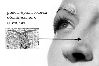 Smells can easily “stir up” our memory and bring back long-forgotten sensations, for example from childhood. The fact is that the centers of the olfactory analyzer are located in humans in the ancient and old cerebral cortex. Next to the olfactory center is the center responsible for our emotions and memory. Therefore, smells are emotionally charged for us, awakening not logical, but emotional memory.
The perception of smell by our olfactory system begins with the nose, or more precisely, with the olfactory epithelium, located in humans in the upper parts of the middle turbinate, in the superior turbinate and the upper part of the nasal septum. The peripheral processes of the receptor cells of the olfactory epithelium end in an olfactory club, decorated with a bunch of microvilli. It is the membrane of these villi (cilia and microvilli) that is the site of interaction between the olfactory cell and the molecules of odorous substances. In humans, the number of olfactory cells reaches 6 million (3 million in each nostril). This is a lot, but in those mammals in whose life the sense of smell plays a significant role, these cells are immeasurably more numerous. For example, a rabbit has about 100 million of them!
In the human embryo, the development of olfactory cells occurs quite quickly. Already in an 11-week fetus they are well differentiated and presumably capable of performing their function.
The receptor cells of the olfactory epithelium are constantly renewed. The life of one cell lasts only a few months or even less. When the olfactory epithelium is damaged, cell regeneration is significantly accelerated.
But how does the excitation of olfactory cells occur? In the last decade, it has become clear that the main role in this process belongs to receptor proteins, whose molecules, interacting with molecules of odorant substances, change their conformation. This leads to the launch of a whole chain of complex reactions, as a result of which the sensory signal is converted into a universal signal from nerve cells. Next, from the receptor cells along their axons, which form the olfactory nerve, the signal is transmitted to the olfactory bulbs. Here its primary processing takes place, and then the signal travels along the olfactory nerve to the brain, where its final analysis occurs.
The ability to perceive odors changes as a person ages. Olfactory acuity reaches its maximum at age 20, stays at the same level for about 30–40 years, and then begins to decline. A particularly noticeable decrease in the acuity of smell occurs in people over 70 years of age, and sometimes even 60 years of age. This phenomenon is called senile hyposmia, or presbiosmia, and is not nearly as harmless as it might seem. Older people gradually cease to perceive the smell of food and therefore lose their appetite. After all, the aroma of food is one of the necessary conditions for the production of digestive juices in the gastrointestinal tract. No wonder they say: “... such a wonderful smell that even my mouth started watering...”. In addition, taste and olfactory perceptions are very close. The odorous substances contained in food products enter the nasal cavity through the nasopharynx, and we smell their aroma. But when we have a runny nose, no matter what we eat, it feels like we are chewing tasteless cardboard. Elderly people with a sharply reduced sense of smell perceive food in the same way. They also lose the ability to determine the quality of food by smell, and therefore can become poisoned by eating low-quality food. It also turns out that older people no longer perceive the smell of mercaptans as unpleasant. Mercaptans are substances added to natural gas used in everyday life (which in itself does not smell like anything from a human point of view) specifically so that its leak can be detected by smell. Old people stop noticing this smell...
But even among young people, sensitivity to the smell of the same substances varies greatly. It also changes depending on environmental factors (temperature, humidity), emotional state and hormonal levels. In pregnant women, for example, against the background of a general decrease in the acuity of smell, sensitivity to certain odors sharply increases. In general, the range of threshold concentrations of various odorous substances perceived by humans is very large - from 10-14 to 10-5 mol per 1 liter of air.
Until now, we have talked mainly about external odors that come from the world around us. But among the odorous substances, there are also those that are released by our body itself and are capable of causing certain behavioral and physiological reactions in other people. Substances with such properties are called pheromones. In the animal world, pheromones play a huge role in the regulation of behavior - we have already written about this in our newspaper (No. 10/1996 and No. 16/1998). Substances have also been discovered in humans that have a certain pheromonal effect during our communication. Such substances are found, for example, in human sweat. In the 70s XX century researcher Martha McClintock discovered that women who live in the same room (for example, in a dormitory) for a long time synchronize their menstrual cycles. And the smell of the secretion of male sweat glands causes the normalization of unstable menstrual cycles in women.

Tapestry “Lady with a Unicorn” – an allegorical depiction of the sense of smell
The smell of the secretion secreted by our axillary sweat glands depends both on the substances secreted by the body itself and on the bacteria present in the sweat glands. After all, it is known that fresh axillary sweat itself (produced profusely, for example, in hot weather) does not have a strong specific odor. But the activity of bacteria contributes to the release of odorous molecules, initially associated with special carrier proteins from the group of lipocaines.
Chemical composition Male and female sweat varies greatly. In women, it is associated with the phases of the menstrual cycle, and a man who has been in intimate relationships with a woman, is able to determine by smell the time of his partner’s ovulation. True, as a rule, this happens unconsciously - it’s just that during this period the girlfriend’s smell becomes the most attractive to him.
In the secretions of the sweat glands of both men and women, in addition to other components, there are two odorous steroids - androstenone (ketone) and androstenol (alcohol). For the first time, these substances were identified as components of the sex pheromone contained in boar saliva. Androstenone has a strong, specific odor, which for many people is similar to the smell of urine. The smell of androstenol is perceived as musky or sandalwood. The content of androstenone and androstenol in men's axillary sweat is much higher than in women's. Studies have shown that the smell of androstenone can affect the physiological and emotional state of people, in particular, suppress the effect of synchronization of sexual cycles described above in women living in the same room. In some situations, the faint smell of androstenone creates a comfortable state of “security” in women, while in men, on the contrary, it causes discomfort and is associated with competition and aggression.
Representatives different cultures can perceive the same odors differently. Such differences were revealed in a completely unique survey conducted in 1986 by National Geographic magazine. The next issue of this magazine included samples of six odorous substances: androstenone, isoamyl acetate (smells like pear essence), galaxolide (smells like synthetic musk), eugenol, a mixture of mercaptans and rose oil. The substances were enclosed in microcapsules applied to paper. When the paper was rubbed with a finger, the capsules were easily destroyed and the smell was released. Readers were asked to smell the proposed substances and then answer the questionnaire. It was necessary to evaluate the intensity of the proposed odors, define them as pleasant, unpleasant or neutral, and talk about the emotions and memories they evoked. Respondents were also asked to indicate their age, gender, occupation, country of residence, race, presence of diseases, etc. For women, it was necessary to indicate the presence of pregnancy. Letters with completed questionnaires came from more than 1.5 million people living on different continents!

Baker of the House of Amun donating incense to Osiris
Many of those who responded did not smell androstenone at all, and the number of people who were not sensitive to this smell varied greatly in different regions globe. So, if in the USA about 30% of women did not feel this smell, then among white women living in Africa there were half as many - about 15%.
We have already talked about the loss of olfactory acuity in older people, which was also clearly revealed during this study. The survey also confirmed that smokers have a much worse sense of smell than non-smokers.
People who, for various reasons, had completely lost their sense of smell also sent their answers to National Geographic. It turns out that there are a lot of such people, including among young people. According to the US National Institutes of Health, in 1969, smell disorders were noted in 2 million people, and by 1981 this figure had increased to 16 million! This situation is largely due to the deterioration of the environmental situation. Among the patients at the Smell and Taste Clinic in Washington, 33% of patients with dysosmia (impaired sense of smell) are people aged 17–20 years. According to researcher Hendricks, in 1988, 1% of the Dutch population had problems with their sense of smell. As for our country, very often people, overwhelmed by other problems, simply do not pay attention to such a “trifle” as a violation or lack of sense of smell. And if they do, they don’t know whether it’s possible in this case health care and where to go for it. Treatment of people with impaired sense of smell is carried out in Moscow, at the ENT clinic of the Moscow Medical Academy. THEM. Sechenov.
What can cause a violation of the sense of smell? Most often, the corresponding disorders are associated with damage to the receptor apparatus of the olfactory analyzer (about 90% of cases), with damage to the olfactory nerve - about 5% of cases, and with damage to the central parts of the brain - the remaining 5% of cases.
The causes of olfactory disorder at the “receptor level” are very diverse and numerous. These include injuries to the olfactory zone and cribriform plate, and inflammatory processes in the nasal cavity, and traumatic brain injuries, and drug intoxication, and allergic reactions, and mutations, and vitamin deficiencies (for vitamins A and B12), and intoxication with heavy metal salts (cadmium , mercury, lead), and inhalation of vapors of irritating substances (formaldehyde), and viral infections (mainly the influenza virus), and ionizing radiation, and much more.
The causes of damage to the olfactory nerve are most often due to infectious diseases, disorders metabolism, toxic effects of drugs, nerve damage during surgery and tumors.
Damage to the centers of the olfactory analyzer can be caused by traumatic brain injury, cerebrovascular accident, brain tumors, genetic and infectious diseases, demyelinating processes, Parkinson's disease, Alzheimer's disease. In the latter two diseases, a decrease in the acuity of smell is often detected in the early stages, which allows treatment to begin earlier.
What is a violation of the sense of smell? It could be complete absence the ability to perceive odors (anosmia) or a decrease in the acuity of smell (hyposmia) of varying degrees of severity. A violation of the sense of smell can also be expressed in the form of a distortion in the perception of odors (aliosmia), in which all odors are perceived “in the same manner.” For example, with cacosmia, all odors seem putrid and fecal; with torsosmia - chemical, bitter, burning or metal odors; with parosmia, “garlic smells like violets.” Mixed cases and phantosmia – olfactory hallucinations – are also possible.
Many of the described smell disorders can be successfully treated, especially if you do not delay visiting a doctor.