Physiology of the peripheral nervous system. Structure of the peripheral part Distinctive features of nerve cells
1. What is the peripheral nervous system? How and where are spinal nerves formed and into what branches are they divided?
The peripheral nervous system is that part of the nervous system that connects the GM and SM with sensitive apparatuses - affectors, as well as with those organs and apparatuses that respond to external and internal stimulation with adaptive reactions (movement, secretion of glands) - effectors.
The PNS consists of:
Nerves (trunks, plexuses, roots)
Nerve nodes
Peripheral endings
Spinal nerves are formed by the fusion of the posterior and anterior rami, which are anatomically and functionally connected to their respective segments of the spinal cord through these branches. Therefore, there are 31 pairs of s/m nerves.
The trunk of the s/m nerve is divided into branches:
· Anterior branch
Posterior branch
Meningeal branch
White connecting thread
2. Posterior branches of the s/m nerves: their zone of innervation and distribution features?
The posterior branch has a segmental structure. Therefore, it innervates areas of the body that have retained segmentation: the deep muscles of the back, neck, and the skin over these areas.
The posterior branches are mixed, divided into lateral and medial branches, their diameter is smaller than the anterior branches. The exceptions are: 1). posterior branch of the 1st cervical s/m nerve (suboccipital nerve) – motor; 2). The posterior branch of the II cervical s/m nerve is sensitive, larger than the anterior one.
3. Anterior branches of the s/m nerves: their zone of innervation and difference from the posterior ones?
The anterior branches are not segmented, they innervate areas of the body that have lost their segmentation, form plexuses, and are a mixed branch.
4. Why do the anterior branches of the s/m nerves form plexuses? The anterior branches of which nerves do not form them? Why?
ANSWER: Plexuses are formed because the anterior branches of the s/m nerves innervate non-segmented areas. Only the anterior branches of the s/m nerves Th2 – Th11 segments retain metamerism; they have a segmental structure; they are called intercostal nerves.
5. What plexuses do you know? Their innervation zone?
Plexus:
· Cervical. From the anterior branches of the 4 upper cervical s/m nerves. Innervates the skin in the neck, diaphragm, and neck muscles.
· Shoulder. Anterior branches of the 4 lower cervical s/m nerves. Innervates the muscles, skin of the upper extremities, superficial muscles of the chest and back.
· Lumbar plexus. Anterior branches of the lumbar nerves. Innervates the skin, muscles of the lower abdomen, thighs.
· Sacral plexus. Formed by sacral nerves
6. Cranial nerves: how do they differ from spinal nerves and what groups are they divided into based on fiber composition?
CN are nerves that arise from the brain. Differences from s/m nerves:
· They do not have a segmental structure, they are different in function, shape, and exit sites.
· Different in fiber composition.
Based on the composition of the fibers, there are 4 groups:
ü Sensitive (1,2,8 pairs of CNs)
ü Motor (3,4,6,11,12 pairs of CN)
ü Mixed (5,7,9,10 pairs of CHN)
ü Having plus vegetative fibers (3,7,9,10 pairs of CN)
7. What are peripheral nerves made of? What connective tissue membranes do they have? What is the perineural space and its significance?
A nerve is a part of the nervous system, which is an elongated cord formed by bundles of nerve fibers and connective tissue membranes.
They have connective tissue membranes of three types:
· Endoneural – m/s with separate nerve fibers, forms separate bundles of nerve fibers;
Perineurium – surrounds several bundles of nerve fibers, formed by two plates:
ü Visceral
ü Parietal
· Epineurium – present in the largest nerves, rich in blood vessels – nourishes the nerve, ensures collateral circulation.
There is a perineural space between the plates, all SN have it, it is debatable in the SMN, it communicates with the subarachnoid space and contains cerebrospinal fluid. Of clinical significance is the movement of the rabies pathogen through this space to the GM and SM.
8. What is a nerve fiber? Their classification according to the caliber and speed of impulses.
A nerve fiber is a process of a nerve cell surrounded by a membrane of lemmocytes.
Based on their caliber and speed, they are divided into:
· Gr.A: thick myelin fibers up to 100 µm, v = 10-120 m/s, form somatic nerves.
· Gr.B: thin myelin fibers 1-3 µm, v=3-14 m/s, form pregangliolar autonomic nerves.
· Gr.S: non-myelinated fibers 0.4-1.2 µm, v=0.6-2.4 m/s, form postgangliolar autonomic nerves (to organs).
9. Intra-trunk structure of nerves.
In addition to the fact that the nerve may include nerve fibers of different types, surrounded by connective tissue membranes and having a perineural space, bundles of nerve fibers can be located in different ways. According to Sinelnikov, they distinguish:
· Cable type (vegetative) – all nerve fibers run parallel;
· Network type (somatic) – an adaptive function, a special form of connections between bundles of nerve fibers.
10. Patterns of location of extraorgan nerves.
· The nerves are paired and diverge symmetrically relative to the central nervous system;
· Nerves reach organs along the shortest path, with the exception of the nerves of those organs that move during their development, while the nerves lengthen and change their path;
· Nerves innervate muscles from those segments that correspond to the myotomes of the muscle anlage; if the muscles move, the nerves lengthen.
· Nerves accompany large arteries and veins, forming neurovascular bundles; they are located in protected places.
11. What determines the types of branching of intraorgan nerves? What types do you know in muscles with different structures and functions?
Options for muscle innervation:
· Main type – small branches from one large nerve;
Any nerve consists of nerve fibers - the conductive apparatus and membranes - the supporting connective tissue frame.
Shells
Adventitium. The adventitium is the densest, fibrous outer membrane.
Epinsvriy. The epineurium is an elastic, elastic connective tissue membrane located under the adventitia.
Perineurium. The perineurium is a covering consisting of 3-10 layers of cells of the epithelioid type, very resistant to stretching, but easily torn when stitched. The perineurium divides the nerve into bundles containing up to 5000-10000 fibers.
Endoneurium. It is a delicate sheath separating single fibers and small bundles. At the same time, it acts as a blood-neural barrier.
Peripheral nerves can be considered as a kind of axonal cables, delimited by more or less complex membranes. These cables are extensions of living cells, and the axons themselves are continually renewed by a flow of molecules. The nerve fibers that make up a nerve are processes from various neurons. Motor fibers are processes of motor neurons of the anterior horns of the spinal cord and nuclei of the brain stem, sensory fibers are dendrites of pseudounstolary neurons of the spinal ganglia, autonomic fibers are axons of neurons of the borderline sympathetic trunk.
A separate nerve fiber consists of the neuron process itself - the axial cylinder and the myelin sheath. The myelin sheath is formed by outgrowths of the Schwann cell membrane and has a phospholipid composition. In this, peripheral nerve fibers differ from the fibers of the central nervous system. where the myelin sheath is formed by processes of oligodendrocytes.
The blood supply to the nerve is carried out segmentally from neighboring tissues or vessels. A longitudinal network of vessels is formed on the surface of the nerve, from which many perforating branches extend to the internal structures of the nerve. With the blood, glucose, oxygen, and low-molecular energy substrates enter the nerve fibers, and decay products are removed.
To perform the function of conducting a nerve fiber, it is necessary to constantly maintain its structure. However, its own structures that carry out biosynthesis are not enough to satisfy the plastic needs in the processes of the neuron. Therefore, the main synthesis occurs in the body of the neuron with subsequent transport of the formed substances along the axon. To a much lesser extent, this the process is carried out by Schwann cells with the further transfer of metabolites to the axial cylinder of the nerve fiber.
Axonal transport.
There are fast and slow types of movement of substances along the fiber.
Fast orthograde axonal transport occurs at a rate of 200-400 mm per day and is mainly responsible for the transport of membrane constituents: phospholigations, lipoproteins and membrane enzymes. Retrograde axonal transport ensures the movement of membrane parts in the opposite direction at a speed of up to 150-300 mm per day and their accumulation around the nucleus in close connection with lysosomes. Slow orthograde axonal transport occurs at a rate of 1-4 mm per day and transports soluble proteins and elements of the internal cellular framework. The volume of substances transported by slow transport is much greater than by fast transport.
Any type of axonal transport is an energy-dependent process performed by contractile proteins analogues of actin and myelin in the presence of macroergs and calcium ions. Energy substrates and ions enter the nerve fiber along with the local blood flow.
Local blood supply to the nerve is an absolutely necessary condition for the implementation of axonal transport.
Neurophysiology of impulse transmission:
The conduction of a nerve impulse along the fiber occurs due to the propagation of a depolarization wave along the sheath of the process. Most peripheral nerves, through their motor and sensory fibers, provide impulse conduction at speeds of up to 50-60 m/sec. The depolarization process itself is quite passive, while the restoration of the resting membrane potential and conductivity is carried out through the functioning of NA/K and Ca pumps. For their work, ATP is required, a prerequisite for the formation of which is the presence of segmental blood flow. Cutting off the blood supply to the nerve immediately blocks the conduction of the nerve impulse.
Semiotics of neuropathies
Clinical symptoms that develop when peripheral nerves are damaged are determined by the functions of the nerve fibers that form the nerve. According to the three groups of fibers, there are three groups of symptoms of suffering: motor, sensory and vegetative.
Clinical manifestations of these disorders may include symptoms of loss of function, which is more common, and symptoms of irritation, the latter being a rarer option.
Motor disorders of the prolapse type are manifested by plegia and paresis of a peripheral nature with low tone, low reflexes and hypotrophy. Symptoms of irritation include convulsive contraction of muscles - cramps. These are paroxysmal, painful contractions of one or more muscles (what we used to call a cramp). Most often, cramps are localized in the mylohyoid muscle, under the occipital muscle, hip adductors, quadriceps femoris, and triceps surae. The mechanism of crampy is not clear enough; partial morphological or functional denervation in combination with autonomic irritation is assumed. In this case, the autonomic fibers take on part of the somatic functions and then the striated muscle begins to respond to acetylcholine similarly to smooth muscles.
Sensory disorders such as prolapse are manifested by hypoesthesia and anesthesia. Symptoms of irritation are more varied: hyperesthesia, hyperpathia (qualitative distortion of sensation with the acquisition of an unpleasant shade), paresthesia (“goosebumps”, burning in the innervation zone), pain along the nerves and roots.
Autonomic disorders are manifested by impaired sweating, suffering in the motor function of hollow internal organs, orthostatic hypotension, and trophic changes in the skin and nails. The irritative variant is accompanied by pain with an extremely unpleasant cutting, twisting component, which occurs mainly when the median and tibial nerves are damaged, as they are the richest in autonomic fibers.
It is necessary to pay attention to the variability of manifestations of neuropathy. Slow changes in the clinical picture that occur over weeks and months actually reflect the dynamics of neuropathy, while changes within hours or one to two days are more often associated with changes in blood flow, temperature, and electrolyte balance.
Pathophysiology of neuropathy
What happens to nerve fibers during nerve diseases?
There are four main options for change.
1. Wallerian degeneration.
2. Axon atrophy and degeneration (axonopathy).
3.Segmental demyelination (myelinopathy).
4.Primary damage to nerve cell bodies (neuronopathy).
Wallerian degeneration occurs as a result of gross local damage to the nerve fiber, often due to mechanical and ischemic factors. The function of conduction through this area of the fiber is completely and immediately impaired. After 12-24 hours, the structure of the axoplasm changes in the distal part of the fiber, but impulse conduction persists for another 5-6 days. On days 3-5, destruction of the nerve endings occurs, and by day 9, they disappear. From 3 to 8 days, the mislin shells are progressively destroyed. In the second week, Schwann cells begin to divide, and by days 10-12 they form longitudinally oriented nerve processes. From days 4 to 14, multiple growth flasks appear on the proximal portions of the fibers. The rate of fiber growth through the s/t at the site of injury can be extremely low, but distally in the uninjured parts of the nerve the regeneration rate can reach 3-4 mm per day. With this type of lesion, good recovery is possible.
Axonal degeneration occurs as a result of metabolic disturbances in the cell bodies of neurons, which then causes disease of the processes. The cause of this condition is systemic metabolic diseases and the action of exogenous toxins. Axonal necrosis is accompanied by the absorption of myelin and remnants of the axial cylinder by Schwann cells and macrophages. The possibility of restoring nerve function in this suffering is extremely low.
Segmental demyelination is manifested by primary damage to the myelin sheaths while the axial cylinder of the fiber is preserved. The severity of the development of disorders may resemble that of mechanical nerve injury, but the dysfunction is easily reversible, sometimes within a few weeks. Pathomorphologically, disproportionately thin myelin sheaths, accumulation of mononuclear phagocytes in the endoneural space, and proliferation of Schwann cell processes around neuronal processes are determined. Functional restoration occurs quickly and in full when the damaging factor ceases.
16-09-2012, 21:50
Description
The peripheral nervous system has the following components:- Ganglia.
- Nerves.
- Nerve endings and specialized sensory organs.
Ganglia
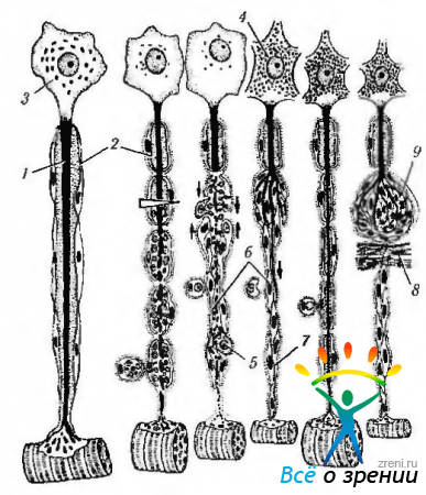
Ganglia are a cluster of neurons that form, in the anatomical sense, small nodules of various sizes, scattered in different parts of the body. There are two types of ganglia - cerebrospinal and autonomic. The bodies of neurons in the spinal ganglia are usually round in shape and vary in size (from 15 to 150 µm). The nucleus is located in the center of the cell and contains distinct round nucleolus(Fig. 1.5.1).

Rice. 1.5.1. Microscopic structure of the intramural ganglion (a) and cytological features of ganglion cells (b): a - groups of ganglion cells surrounded by fibrous connective tissue. On the outside, the ganglion is covered with a capsule to which adipose tissue is adjacent; b-neurons of the ganglion (1 - inclusion in the cytoplasm of the ganglion cell; 2 - hypertrophied nucleolus; 3 - satellite cells)
Each neuron body is separated from the surrounding connective tissue by a layer of flattened capsular cells (amphicytes). They can be classified as cells of the glial system. The proximal process of each ganglion cell in the dorsal root divides into two branches. One of them flows into the spinal nerve, in which it passes to the receptor ending. The second enters the dorsal root and reaches the posterior column of gray matter on the same side of the spinal cord.
Ganglia of the autonomic nervous system similar in structure to the cerebrospinal ganglia. The most significant difference is that the neurons of the autonomic ganglia are multipolar. In the orbital area, various autonomic ganglia are found that provide innervation to the eyeball.
Peripheral nerves
Peripheral nerves are clearly defined anatomical structures and are quite durable. The nerve trunk is enveloped from the outside in a connective tissue sheath along its entire length. This outer sheath is called the epinervium. Groups of several bundles of nerve fibers are surrounded by perineurium. Strands of loose fibrous connective tissue surrounding individual bundles of nerve fibers are separated from the perineurium. This is the endoneurium (Fig. 1.5.2).

Rice. 1.5.2. Features of the microscopic structure of the peripheral nerve (longitudinal section): 1- axons of neurons: 2- nuclei of Schwann cells (lemmocytes); 3-Ranvier interception
Peripheral nerves are abundantly supplied with blood vessels.
The peripheral nerve consists of varying numbers of densely packed nerve fibers, which are the cytoplasmic processes of neurons. Each peripheral nerve fiber is covered with a thin layer of cytoplasm - neurilemma, or Schwann's membrane. The Schwann cells (lemmocytes) involved in the formation of this membrane are derived from neural crest cells.
In some nerves, between the nerve fiber and the Schwann cell there is myelin layer. The former are called myelinated, and the latter - unmyelinated nerve fibers.
Myelin(Fig. 1.5.3)

Rice. 1.5.3. Peripheral nerve. Ranvier interceptions: a - light optical microscopy. The arrow indicates the interception of Ranvier; b-ultrastructural features (1-axoplasm of the axon; 2-axolemma; 3 - basement membrane; 4 - cytoplasm of the lemmocyte (Schwann cell); 5 - cytoplasmic membrane of the lemmocyte; 6 - mitochondrion; 7 - myelin sheath; 8 - neurofilaments; 9 - neurotubules ; 10 - nodular interception zone; 11 - plasmalemma of the lemmocyte; 12 - space between neighboring lemmocytes)
does not cover the nerve fiber completely, but is interrupted after a certain distance. Sites of myelin interruption are designated nodes of Ranvier. The distance between successive nodes of Ranvier varies from 0.3 to 1.5 mm. Nodes of Ranvier are also present in the fibers of the central nervous system, where myelin forms oligodendrocytes (see above). Nerve fibers branch precisely at the nodes of Ranvier.
How is the myelin sheath of peripheral nerves formed?? Initially, the Schwann cell wraps around the axon so that it lies in the groove. Then this cell is wound around the axon. In this case, sections of the cytoplasmic membrane along the edges of the groove come into contact with each other. Both parts of the cytoplasmic membrane remain connected, and the cell is then seen to continue to spiral around the axon. Each turn in a cross section has the appearance of a ring consisting of two lines of the cytoplasmic membrane. As coiling proceeds, the cytoplasm of the Schwann cell is squeezed into the cell body.
Some afferent and autonomic nerve fibers do not have a myelin sheath. However, they are protected by Schwann cells. This occurs due to the pressing of axons into the body of Schwann cells.
The mechanism of nerve impulse transmission in unmyelinated fiber is covered in physiology manuals. Here we will only briefly describe the main principles of the process.
It is known that the cytoplasmic membrane of the neuron is polarized, i.e. between the inner and outer surfaces of the membrane there is an electrostatic potential equal to - 70 mV. Moreover, the inner surface has a negative charge, and the outer surface has a positive charge. This state is ensured by the action of the sodium-potassium pump and the peculiarities of the protein composition of the intracytoplasmic contents (the predominance of negatively charged proteins). The polarized state is called the resting potential.
When stimulating a cell, i.e. irritating the cytoplasmic membrane with a wide variety of physical, chemical and other factors, Initially, depolarization occurs, and then repolarization of the membrane. In the physicochemical sense, this results in a reversible change in the concentration of K and Na ions in the cytoplasm. The repolarization process is active using energy reserves of ATP.
A wave of depolarization - repolarization propagates along the cytoplasmic membrane (action potential). Thus, the transmission of a nerve impulse is nothing more than propagating action potential wave I.
What is the significance of the myelin sheath in the transmission of nerve impulses? It is stated above that myelin is interrupted at the nodes of Ranvier. Since only at the nodes of Ranvier does the cytoplasmic membrane of the nerve fiber come into contact with tissue fluid, only in these places is it possible for the membrane to depolarize in the same way as in unmyelinated fibers. Throughout the rest of the process, this process is impossible due to the insulating properties of myelin. As a result, between the nodes of Ranvier (from one area of possible depolarization to another), the transmission of a nerve impulse carried out by intracytoplasmic local currents. Because the electrical current travels much faster than a continuous wave of depolarization, transmission of a nerve impulse in a myelinated nerve fiber occurs much faster (50 times), and the speed increases with increasing nerve fiber diameter, due to a decrease in internal resistance. This type of nerve impulse transmission is called saltatory. i.e. jumping. Based on the above, the important biological significance of myelin sheaths is evident.
Nerve endings
Afferent (sensitive) nerve endings (Fig. 1.5.5, 1.5.6).

Rice. 1.5.5. Features of the structure of various receptor endings: a - free nerve endings; b- Meissner's body; c - Krause flask; d - Vater-Pacini body; d - Ruffini body

Rice. 1.5.6. The structure of the neuromuscular spindle: a-motor innervation of intrafusal and extrafusal muscle fibers; b spiral afferent nerve endings around intrafusal muscle fibers in the area of nuclear bags (1 - neuromuscular effector endings of extrafusal muscle fibers; 2 - motor plaques of intrafusal muscle fibers; 3 - connective tissue capsule; 4 - nuclear bag; 5 - sensitive ring-spiral nerve endings around nuclear bags; 6 - skeletal muscle fibers; 7 - nerve)
Afferent nerve endings They are the terminal apparatus of the dendrites of sensitive neurons, located everywhere in all human organs and providing information to the central nervous system about their condition. They perceive irritations emanating from the external environment, converting them into a nerve impulse. The mechanism of occurrence of a nerve impulse is characterized by the already described phenomena of polarization and depolarization of the cytoplasmic membrane of the nerve cell process.
Exists a number of classifications of afferent endings- depending on the specificity of stimulation (chemoreceptors, baroreceptors, mechanoreceptors, thermoreceptors, etc.), on structural features (free and non-free nerve endings).
Olfactory, taste, visual and auditory receptors, as well as receptors that perceive the movement of body parts relative to the direction of gravity, are called special sense organs. In subsequent chapters of this book we will dwell in detail only on visual receptors.
Receptors vary in shape, structure and function. In this section, our task is not to describe in detail the various receptors. Let us mention only a few of them in the context of describing the basic principles of the structure. In this case, it is necessary to point out the differences between free and non-free nerve endings. The first are characterized by the fact that they consist only of the branching of the axial cylinders of the nerve fiber and glial cells. At the same time, they contact the branches of the axial cylinder with the cells that excite them (receptors of epithelial tissues). Non-free nerve endings are distinguished by the fact that they contain all the components of a nerve fiber. If they are covered with a connective tissue capsule, they are called encapsulated(Vater-Pacini corpuscle, tactile Meissner corpuscle, Krause flask thermoreceptors, Ruffini corpuscle, etc.).
The structure of muscle tissue receptors is varied, some of which are found in the external muscles of the eye. In this regard, we will dwell on them in more detail. The most common receptor in muscle tissue is neuromuscular spindle(Fig. 1.5.6). This formation records the stretching of the fibers of the striated muscles. They are complex encapsulated nerve endings that have both sensory and motor innervation. The number of spindles in a muscle depends on its function and the higher the more precise movements it has. The neuromuscular spindle is located along the muscle fibers. The spindle is covered with a thin connective tissue capsule (a continuation of the perineurium), inside which there are thin striated intrafusal muscle fibers two types:
- fibers with a nuclear bag - the expanded central part of which contains clusters of nuclei (1-4 fibers/spindle);
- fibers with a nuclear chain - thinner with nuclei arranged in the form of a chain in the central part (up to 10 fibers/spindle).
Sensory nerve fibers form ring-spiral endings on the central part of intrafusal fibers of both types and cluster-shaped endings at the edges of fibers with a nuclear chain.
Motor nerve fibers- thin, form small neuromuscular synapses along the edges of intrafusal fibers, ensuring their tone.
Muscle stretch receptors are also neurotendon spindles(Golgi tendon organs). These are spindle-shaped encapsulated structures about 0.5-1.0 mm long. They are located in the area where the fibers of the striated muscles connect with the collagen fibers of the tendons. Each spindle is formed by a capsule of flat fibrocytes (a continuation of the perineurium), which encloses a group of tendon bundles entwined with numerous terminal branches of nerve fibers, partially covered with lemmocytes. Excitation of the receptors occurs when the tendon is stretched during muscle contraction.
Efferent nerve endings carry information from the central nervous system to the executive organ. These are the endings of nerve fibers on muscle cells, glands, etc. A more detailed description of them will be given in the relevant sections. Here we will dwell in detail only on the neuromuscular synapse (motor plaque). The motor plaque is located on the fibers of the striated muscles. It consists of the terminal branching of the axon, forming the presynaptic part, a specialized area on the muscle fiber corresponding to the postsynaptic part, and the synaptic cleft separating them. In large muscles, one axon innervates a large number of muscle fibers, and in small muscles (extrinsic muscles of the eye), each muscle fiber or a small group of them is innervated by one axon. One motor neuron, together with the muscle fibers it innervates, forms a motor unit.
The presynaptic part is formed as follows. Near the muscle fiber, the axon loses its myelin sheath and gives rise to several branches, which are covered on top with flattened lemmocytes and a basement membrane that passes from the muscle fiber. The axon terminals contain mitochondria and synaptic vesicles containing acetylcholine.
The synaptic cleft is 50 nm wide. It is located between the plasma membrane of the axon and muscle fiber branches. It contains basement membrane material and processes of glial cells that separate adjacent active zones of one end.
Postsynaptic part It is represented by a muscle fiber membrane (sarcolemma), forming numerous folds (secondary synaptic clefts). These folds increase the total area of the gap and are filled with material that is a continuation of the basement membrane. In the area of the neuromuscular ending, the muscle fiber does not have striations. contains numerous mitochondria, cisterns of rough endoplasmic reticulum and a cluster of nuclei.
The mechanism of transmission of nerve impulses to muscle fibers similar to that in a chemical interneuron synapse. When the presynaptic membrane is depolarized, acetylcholine is released into the synaptic cleft. The binding of acetylcholine to cholinergic receptors in the postsynaptic membrane causes its depolarization and subsequent contraction of the muscle fiber. The mediator is cleaved from the receptor and quickly destroyed by acetylcholinesterase.
Peripheral nerve regeneration
When a section of a peripheral nerve is destroyed within a week, ascending degeneration of the proximal (closest to the neuron body) part of the axon occurs, followed by necrosis of both the axon and the Schwann sheath. An extension (retraction flask) is formed at the end of the axon. In the distal part of the fiber after its transection, descending degeneration is observed with complete destruction of the axon, disintegration of myelin and subsequent phagocytosis of detritus by macrophages and glia (Fig. 1.5.8).
Rice. 1.5.8. Regeneration of myelinated nerve fiber: a - after cutting the nerve fiber, the proximal part of the axon (1) undergoes ascending degeneration, the myelin sheath (2) in the area of damage disintegrates, the perikaryon (3) of the neuron swells, the nucleus shifts to the periphery, the chromaphilic substance (4) disintegrates; b-distal part, associated with the innervated organ, undergoes descending degeneration with complete destruction of the axon, disintegration of the myelin sheath and phagocytosis of detritus by macrophages (5) and glia; c - lemmocytes (6) are preserved and mitotically divide, forming strands - Bugner's ribbons (7), connecting with similar formations in the proximal part of the fiber (thin arrows). After 4-6 weeks, the structure and function of the neuron is restored, thin branches grow distally from the proximal part of the axon (thick arrow), growing along the Buegner strip; d - as a result of regeneration of the nerve fiber, the connection with the target organ is restored and its atrophy regresses: e - when an obstacle occurs (8) in the path of the regenerating axon, the components of the nerve fiber form a traumatic neuroma (9), which consists of growing branches of the axon and lemmocytes
The beginning of regeneration is characterized first by proliferation of Schwann cells, their movement along the disintegrated fiber with the formation of a cellular cord lying in the endoneurial tubes. Thus, Schwann cells restore structural integrity at the incision site. Fibroblasts also proliferate, but more slowly than Schwann cells. This process of proliferation of Schwann cells is accompanied by the simultaneous activation of macrophages, which initially capture and then lyse the material remaining as a result of nerve destruction.
The next stage is characterized growth of axons into clefts, formed by Schwann cells, pushing from the proximal end of the nerve to the distal one. At the same time, thin branches (growth cones) begin to grow from the retraction flask towards the distal part of the fiber. The regenerating axon grows in the distal direction at a speed of 3-4 mm per day along ribbons of Schwann cells (Bugner's ribbons), which play a guiding role. Subsequently, differentiation of Schwann cells occurs with the formation of myelin and surrounding connective tissue. Axon collaterals and terminals are restored within several months. Nerve regeneration occurs only if there is no damage to the neuron body, a small distance between the damaged ends of the nerve, the absence of connective tissue between them. When an obstacle occurs in the path of the regenerating axon, an amputation neuroma develops. There is no regeneration of nerve fibers in the central nervous system.
Article from the book: .
Each peripheral nerve consists of a large number of nerves
fibers united by connective tissue membranes (Fig. 265- A).
In a nerve fiber, regardless of its nature and functional purpose,
definitions, distinguish between “throat cylinder- cylindroaxis, covered with its own
sheath - axolemma -^ and nerve sheath - neurolemma. When on-
in the presence of a fat-like substance - myelin - nerve fiber
called pulpy or myelin-*■ neurofibra myelinate, and with it"
absence - pulpless or amyelin- neurofibra amyelinata (go-
long nerve fibers - neurofibria nuda).
The significance of the pulpy shell is that it contributes to
better conduction of nervous stimulation. In the pulpless nerve fibers
excitation is carried out at a speed of 0.5-2 m/s, while in the soft
cat fibers - 60-120 m/s". The diameter of individual nerve fibers
are divided into thick pulpy ones (from 16-26 microns in horses, ruminants
up to 10-22 microns in a dog)>-efferent somatic; medium pulpy
(from 8-15 microns in horses, ruminants to 6-^-8 microns in dogs) - afferent
somatic; thin (4-8 microns) - efferent vegetative (Fig. 265- B).
Non-pulp nerve fibers are part of both somatic and
and visceral nerves, but in quantitative terms there are more of them in the vega-
tative nerves. They differ in both the diameter and shape of the kernels
neurolemmas: 1) small-fleshed, or non-fleshed, fibers with a rounded
shape of the cores (fiber diameter 4-2.5 microns, core size 8X4.6 microns, dis-
distance between cores 226t-345 microns); 2) low-pulp or pulpless
fibers with an oval-elongated shape of the neurolemma nuclei (fiber diameter
1-2.5 microns, core size 12.8 X 4 microns, distance between cores 85-
180 µm); 3) non-pulp fibers with spindle-shaped nuclei neurosis
lemmas (fiber diameter 0.5-1.5 µm, core size 12.8 x 1.2 µm, dis-
Fig-265. Structure of the peripheral nerve!
A- nerve on a transverse section: 1
- epineurium; 2
- perineurium; 3
- endoneurium!
4
- neurofibra myelinata; 5
- cylindrix; B- composition of nervous fibers in somatic
sheep's nerve; 1, 2, 3
- neurofibra myelinata; 4
- neurofibra amyelinata; 5,
6,7 -
neurofibra nuda; a- lemmocytus; n- incisio myelini; O- isthmus nodi.
distance between fibers is 60-120 microns). In animals of different species these are,
indicators may not be the same.
Nerve sheaths. Nerve fibers extending from the brain through
connective tissue are combined into bundles that form the basis of the peri-
spherical nerves. In each nerve, connective tissue elements are involved
occur in the formation of: a) inside the fascicular base - endoneurium, located
existing in the form of loose connective tissue between individual nerves
fibers; b) connective tissue membrane covering individual
groups of nerve fibers, or perineurium- perineurium. In this shell
on the outside there is a double layer of flat epithelial cells ependi-
of a grave nature, which form around the nerve bundle of the perineum
vaginal vagina, or perineural space-spatium peri-
neurii. 0t basilar inner layer of the perineural lining
connective tissue fibers extend deep into the nerve bundle,
forming intrafascicular perineural septa-septum peri-
neurii; the latter serve as a place for the passage of blood vessels, as well as
also participate in the formation of the endoneurium. > .
Perineural sheaths accompany bundles of nerve fibers on
along their entire length and are divided as the nerve divides into smaller branches.
The cavity of the perineural vagina communicates with the subarachnoid
and subdural spaces of the spinal cord or brain and contain-
lives a small amount of cerebrospinal fluid (neurogenic route of penetration of vi-
rus of rabies into the central parts of the nervous system).
Groups of primary nerve bundles through dense unformed
of connective tissue are combined into larger secondary and
tertiary bundles of nerve trunks and form the external connection in them
calf shell, izhepineurium- epineurium. In epineurium in comparison
Larger blood vessels and lymphatics pass through the endoneurium
Chinese vessels - vasa nervorum. Around the nerve trunks there is one or another
quantity (depending on the location of passage) of loose connective tissue
tissue that forms an additional periphery of the nerve trunk
Nervous (protective) sheath - paraneural i.e. in the immediate vicinity
In spite of the nerve bundles, it is transformed into the epineural sheath.
Date added: 2015-08-06 | Views: 379 | Copyright infringement
| | | | | | | | | | | | | | | | | | | | | | | | | | | | | | | | | | |
CONCEPT OF THE PERIPHERAL NERVOUS SYSTEM
TRAINING MODULE 7. FUNCTIONAL ANATOMY OF THE PERIPHERAL NERVOUS SYSTEM
LEARNING OBJECTIVES
AFTER STUDYING THE MODULE, THE STUDENT SHOULD:
HAVE AN INTRODUCTION ABOUT: the structures of the peripheral nervous system; the importance of the peripheral nervous system in the transmission of information; the principle of formation of sensory, motor and parasympathetic fibers of the cranial nerves; main nuclei of cranial nerves.
KNOW: the structure of the spinal nerves, their number; branches of the spinal nerves; structure and features of innervation of the posterior branches of the spinal nerves; plexus of the anterior branches of the spinal nerves, zones of their innervation; names and functional types of XII pairs of cranial nerves; formation, exit points from the cranial cavity, areas of innervation of cranial nerves.
BE ABLE: show the main nerves of the somatic plexuses of the anterior branches of the spinal nerves and 12 pairs of cranial nerves on models and tables; show the zones of innervation of the spinal and cranial nerves in the atlas, on tables and models.
THEORETICAL PART
The peripheral nervous system is that part of the nervous system that is located outside the brain and spinal cord. Through the peripheral part of the central nervous system it regulates the functions of all organs and systems. The peripheral nervous system includes the spinal and cranial nerves, their sensory nodes, nerves, nodes and plexuses of the autonomic nervous system, receptors and effectors.
Depending on the part of the central nervous system from which the peripheral nerves arise, there are spinal nerves (SCN), which arise from the spinal cord, and cranial (cranial) nerves (CN), which arise from the brain stem. Thanks to the spinal nerves, motor and sensory somatic innervation of the torso, limbs and part of the neck is carried out, as well as autonomic innervation of internal organs. Cranial nerves innervate the head and partly the neck.
A bundle of nerve fibers forms a nerve (nerve trunk), surrounded by a connective tissue sheath. The nerve usually includes a large number of motor, sensory, and sometimes autonomic fibers that innervate various tissues and organs. Such nerves are called mixed. There are also purely motor, sensory and autonomic (parasympathetic) nerves.
There are nerves (branches) cutaneous, sensory, superficial - muscular and motor - deep. Cutaneous nerves are located in the subcutaneous fat layer. They contain sensitive somatic fibers that innervate the skin and autonomic fibers that innervate the sebaceous and sweat glands, blood vessels and muscles that lift the hair. Muscle nerves are usually part of neurovascular bundles, located deep between muscles and contain motor, sensory and autonomic nerve fibers that innervate skeletal muscles, joints, bones, blood vessels and internal organs.
Motor nerves are formed by the axons of motor neurons of the anterior horns of the spinal cord and the motor nuclei of the cranial nerve. Sensory nerves are formed by processes of afferent neurons of the spinal and cranial nodes (ganglia). Autonomic nerves consist of processes of neurons of the lateral horns of the spinal cord and autonomic nuclei of the cranial nerve. They are prenodular nerve fibers and follow to the autonomic nerve ganglia and plexuses. Postnodal fibers extend from these nodes and plexuses further to the internal organs and tissues. Vegetative fibers are part of the majority of the spinal nerves and all the spinal nerves.
Large nerves often enter neurovascular bundles (highways), surrounded by a common connective tissue sheath. The composition of such a bundle usually includes an artery, veins, lymphatic vessels, and a nerve.