Inguinal hernia: symptoms, diagnosis, treatment. Congenital and sliding inguinal hernias. Diagnostics. Features of surgical treatment of hernia of the white line of the abdomen
Sliding we should call those inguinal hernias in which in the formation of the hernial sac, in addition to the parietal, the visceral peritoneum also takes part, covering a small area of the adjacent slipped organ, and its other parts, devoid of serosa, are located extraperitoneally, outside the sac, in the retroperitoneal or preperitoneal tissue. In more rare cases, the visceral peritoneum covers an almost completely slipped organ, which protrudes and hangs into the lumen of the sac, or in very rare cases, the hernial sac is absent, and the entire protrusion is formed only by those segments of the slipped organ that are almost not covered by peritoneum.
Sliding hernias, or “sliding hernias,” can occur in all forms of inguinal hernias. The first to propose the term “landslide” was Mitchell Banks (1887). There are mainly sliding inguinal hernias of the colon, bladder and internal female genital organs.
Sliding hernias of the colon and female genital organs are much more common with oblique inguinal hernias, and of the bladder - with direct and supravesical hernias, since they are located closer to the middle and supravesical fossae. A distinctive feature of sliding hernias is that the colon, bladder and internal female genital organs, under the influence of various reasons, sometimes starting from the embryonic period, can gradually, with some of their sections not covered by peritoneum, descend or, as it were, slide along loose retroperitoneal or preperitoneal tissue to the internal hernial orifice, exit the abdominal cavity and become an integral part of the hernial protrusion and hernial sac to one degree or another. Along with this, free loops of intestines, omentum and other internal organs can enter the cavity of the hernial sac, as with a normal hernia, contributing to its further enlargement.
The formation of sliding inguinal hernias can involve the right and left bladder, ovaries, tubes, and uterus; in addition, on the right - the cecum and the appendix or only the appendix, the ascending colon, very rarely - the terminal part of the small intestine; on the left - the sigmoid colon, the descending colon, and in some cases with large anomalies of location and with a common mesentery (mesenterium commune) of the large and small intestines - the cecum with the appendix or only the appendix. Slipping of other organs - ureters, kidneys - is extremely rare.
Sliding inguinal hernia of the colon
For the first time (according to Iason) Galen mentioned this kind of hernia; Rousteus (1631), Spigelius (1645) also mentioned them.
Most authors believe that the first more detailed description of such hernias of the cecum was given by J. Otto (1688): the hernia of the cecum was mistaken for a hydrocele and opened during surgery; the patient died. The first mention of hernias of the sigmoid colon belongs to N. Pott (1783). Based on his anatomical studies, Scarpa (1812) described similar hernias of the large intestines in more depth, proposing to distinguish between two forms: congenital and acquired.
To understand a number of conditions for the formation of sliding inguinal hernias and the topography of the colon, it is necessary to take into account some embryological data. During the period of embryonic life, as is known, the homogeneous primary intestinal tube, located almost vertically, makes its complex development path intraperitoneally on the common mesentery. At 4-6 weeks, the first rudiment of the initial part of the large intestine appears - the cecum in the form of a small diverticulum, located almost under the liver, and then gradually descending. In a 12-week embryo, when the intestinal rotation ends, the cecum is located at the level of the anterior superior iliac spine. The mesentery of the colon remains only at the mobile transverse colon and the lower part of the sigmoid colon. In other sections - the ascending, descending colon and the upper part of the sigmoid colon, the peritoneum is welded to one degree or another over varying lengths and fuses with the posterolateral abdominal wall. In rare cases, if the development is abnormal, this primary fusion does not occur, and the common mesentery of the colon along its entire length remains mobile.
Thus, on the left, all three sections of the colon can participate in sliding to one degree or another. A.Yu. Sozon-Yaroshevich, based on studies on corpses of various ages (from embryos to 70 years old), expressed the opinion that with a narrow male pelvis, the sigmoid colon, being more vertical, slides out more easily. Due to the fact that A.Yu. Sozon-Yaroshevich, with a narrow pelvis, more often found a high triangular shape of the inguinal space with a large muscle defect; he explained why men are more likely to have inguinal hernias and, in particular, sliding ones. He also noted that with age, when significant ptosis develops, both the descending colon and, rarely, the colon pelvinum can participate in the formation of sliding hernias.
The study of the etiology and pathogenesis of sliding hernias of the colon led to their division into:
1) congenital, when various anomalies of the development and location of the large intestines can be detected very early or less often - appear later in the future;
2) acquired when the sliding of these organs occurs gradually during the formation of ordinary inguinal hernias.
The theory of the congenital formation of sliding hernias connects them with the process of descent of the testicle into the scrotum (Scarpa, Baumgartner, Lockwood, Fleisig, etc.). It is known that in embryos 2-3 months of uterine life, the testicles are located at the level of 2-3 lumbar vertebrae retroperitoneally on the posterior wall of the abdomen, from them to the future inguinal canals, inguinal cords covered with peritoneum descend. The testicle has its own mesentery, where a vascular fold is formed. On the right side, this fold, located in close proximity to the cecum, can be fused with it. As it descends, the testicle pulls the cecum with it. With this, some authors wanted to explain why cecal hernias are somewhat more common. During surgical interventions, some authors found similar relationships (Baumgartner, E.D. Dmitrieva, etc.). On the other hand, Vernon (1923) in 3 operated and thoroughly examined cases found neither adhesions nor a pronounced vascular fold. Obviously, these formations should be considered unstable, and Lockwood considers them a developmental defect. Not all authors have predominant right-sided sliding hernias of the cecum. According to E.A. Ryan, there were 4.5 times fewer right-sided sliding hernias than left-sided ones. The formation of sliding inguinal hernias of the cecum and sigmoid colon from an abnormally very low location of the large intestine should also be attributed to congenital origin, standing outside the process of descent of the testicle.
Acquired sliding hernias have a different explanation and, according to their origin, are divided into primary and secondary. Some authors (Erces, I.L. Tsimkhes, E.D. Dmitrieva, A.V. Ilyashenko, etc.) suggest that with coprostasis, frequent overflow and distension of the large intestines with gases and feces, softening of the intestinal wall sometimes occurs, and, if the peritoneum is more loosely fused to the intestinal wall, partial detachment may occur. The intestine seems to slip out, emerge from under the serosa, and in the future it can move, slide in loose retroperitoneal tissue, and the detached layer of the mesentery, turning out, forms a hernial sac (Sprengel).
Therefore, such hernias, in which from the very beginning the sliding of the wall in its various segments, devoid of serosa, is the primary, initial moment of the formation of a hernial protrusion, are called primary acquired sliding hernias.
Secondary acquired sliding hernias, the origin of which is clear, are considered to be those when, with an enlarging ordinary inguinal hernia, a mechanical, secondary, gradual contraction by the peritoneum of the immediately adjacent segments of the intestinal wall devoid of serosa occurs.
We must admit, together with the majority of authors (N.M. Tereshenkov and S.P. Fedorov, A.V. Gizhitsky, E.D. Dmitrieva, N.V. Antelava, A. Sozon-Yaroshevich, P.S. Kakhidze and M .I. Pototsky) that the formation of so-called secondary sliding hernias occurs much more often, especially with periperitoneal sliding hernias. We must also agree with Fleissig that the theory of secondary acquired sliding hernias can be accepted for cases where there is a large hernial sac in periperitoneal hernias, but not for those when there is a small hernial sac and the extraperitoneal segment of the colon descends in front. In such cases, one should rather think about either the congenital origin of sliding hernias, or primary acquired hernias.
According to E.A. Ryan, especially with relatively large sliding inguinal hernias, often during hernia repair, upon careful examination, a vaginal cord was found near the hernial sac in the elements of the spermatic cord; as emphasized by E.A. Ryan, this proves that in most cases, sliding indirect inguinal hernias of the colon are acquired and not congenital.
It must be assumed that in the overall complex complex of development of sliding hernias, in most cases the combination of all those many factors that were discussed in Chapter IV about the etiology and pathogenesis of hernias plays a role. Baumgartner (1905) in his dissertation proposed dividing sliding hernias of the colon into three groups depending on the location and relationship of the hernial sac and the serous layer of the intestinal wall.
1) Intraperitoneal, intraperitoneal hernias (herniae intraperitoneales) with a complete hernial sac. In these forms, the parietal peritoneum and part of the intestine, which is entirely covered with serosa with a very short mesentery, take part in the formation of the hernial sac; located in close proximity to the hernial orifice and neck, it seems to hang into the cavity of the sac. The vessels supplying the colon enter through this very short mesentery.
2) Hernias are paraperitoneal, periperitoneal (herniae paraperitoneales), hernias with a complete or incomplete sac, in the formation of which the parietal peritoneum and partly the serosa covering the large intestine in this segment take part. Segments of the colon, devoid of serous cover along its posterior surface, participate in the hernial protrusion and come into direct contact with the retroperitoneal tissue, with tissue in the inguinal canal, scrotum and spermatic cord. The feeding vessels approach through the retroperitoneal tissue and can be easily damaged during surgery.
3) Extraperitoneal hernias, extraperitoneal hernias (herniae extraperitoneales), hernias without a sac. Hernial protrusion involves only segments of the colon that are free from serous cover. Located in the retroperitoneal tissue, they descend through the hernial orifice and can be located in the inguinal canal and below, next to the spermatic cord. The vessels approach through the retroperitoneal tissue and can also be easily damaged by bowel leakage. With this type of sliding hernia, it is very easy to mistake the intestinal wall for a hernial sac and damage it. This rare group, as not having a hernial sac, is conventionally designated by some authors as a hernia.
Some American surgeons, such as E.A. Ryan, intraperitoneal sliding hernias are called intrasaccular, paraperitoneal - peri-saccular, extraperitoneal - extra-saccular, and this also well expresses the relationship of the slipped intestinal wall with the hernial sac (see Appendix No. 4).
This has a certain practical significance when choosing a method of surgical treatment. In the latter form, when the bag is small, it is easier to damage the intestinal wall and the vessels feeding it; with a large bag it is easier to use the method of peritonization of the intestinal wall and mesentery.
As for sliding hernias of the cecum, Froilich and A.V. Ilyashenko, adhering to the Baumgart-peg classification, defines them as follows. Intraperitoneal hernias of the cecum can be recognized as sliding only when the intestine is either directly in the hernial orifice, or slightly lower and protrudes anteriorly; therefore, in most cases they cannot be completely reduced. Hernias should be recognized as ordinary when the cecum, with its great mobility (coecum mobile), forms the free contents of the hernial sac and is easily reduced into the abdominal cavity. Such cases are not so rare and, according to statistics from I.M. Derevianko (1954), occur in 2.4% of all inguinal hernias. This must be remembered in order to take into account only the true forms of sliding hernias of the cecum and not artificially increase their number. For example, Hilgenreiner (1910) discovered 22 cecal hernias and recognized only 8 of them as sliding ones.
Paraperitoneal hernias are those in which the cecum, with more or less of its posterior surface, devoid of serosa, lies outside the hernial sac.
Extraperitoneal hernias are considered to be those in which there is no hernial sac, almost half or more of the surface of the cecum is not covered with serosa, and this part is located in the hernial protrusion.
According to Tuffier, in a study of 120 adult cadavers, a cecum was found in 9 cases, partially not covered by peritoneum on the posterior wall. On the other hand, Cavailon and Leriche (1907), based on their anatomical studies, denied the possibility of extraperitoneal sliding hernias of the cecum. In all the cases they studied, the cecum was covered with serosa to one degree or another, so they believed that hernias of the cecum could only be intraleritoneal.
The most common type is peritoneal sliding hernia of the colon. According to E.A. Ryan, of 313 sliding oblique inguinal hernias of large intestines, 95% were peripacicular (peri-peritoneal), intra-sac (intraperitoneal) - 4.7%, and extra-sac (extraperitoneal) - 0.3% (1 case). Sliding hernias with a large sac are much more common; according to A.V. Ilyashenko, out of 76 operated sliding hernias of the colon, 71 were periperitoneal, and 5 were intraperitoneal, of which 61 were with a large hernial sac and 15 were with a small hernial sac; There were no extraperitoneal hernias.
Sliding inguinal hernias of the colon in children are relatively rare. S.Ya. Doletsky cites 1 case of a sliding hernia of the sigmoid colon. Goldstein and Potts (1958), out of 44 cases of sliding inguinal hernias in girls, did not find a single case of colon hernia in children under 10 years of age. According to N.I. Romantsev (1935), out of 27 sliding hernias of the colon in children, there were 26 sliding hernias of the appendix and cecum and 1 of the sigmoid colon.
Inflammatory phenomena in sliding hernias can lead to adhesions in the hernial sac and to cicatricial changes in the retroperitoneal tissue and surrounding tissues. Scars can also involve feeding vessels, which can cause significant difficulties during surgery.
In sliding hernias of the colon, the cecum and appendix are strangulated more often than the sigmoid colon. Incarceration of the cecum with the appendix does not necessarily lead to inflammatory changes in the appendix. There is no clear division in the statistics of hernial appendicitis with strangulation of sliding and non-sliding hernias of the cecum and appendix, since no significant difference is observed in the course.
The vermiform appendix can be located in sliding hernias of the cecum and appendix, either inside the sac and become inflamed there, or outside the sac (partially or almost completely) and become inflamed outside the sac. Such rare cases were described by E.M. Dmitrieva et al. During herniotomy of a right-sided oblique inguinal sliding hernia in a 60-year-old patient, we observed a case of almost complete extra-abdominal presence of a non-inflamed appendix, located within the scrotum next to the hernial sac. In such cases, inflammation of the appendix will occur outside the sac, and therefore signs of inflammation of the scrotum (swelling, redness) will be more pronounced.
Cases of intussusception of the cecum and small intestine into the ascending intestine during sliding hernias have been described; A.V. Gizycki cites the observations of Bennet and Demeau, although these cases are doubtful, since they are unlikely to occur with a fixed cecum in sliding hernias. Other complications due to long-term large displacements of the intestines may develop disorders of the gastrointestinal tract of various types with symptoms of flatulence, bloating and constipation. Due to congestion from prolonged pressure, testicular atrophy may occur, as well as hydrocele and decreased sexual function.
Sliding inguinal hernias of the colon are mainly found in men. According to A.P. Krymov, the ratio of men to women is 16:1, according to P.S. Kakhidze - 26: 1; by E.A. Ryan, all 313 cases of indirect sliding inguinal hernia were in men. Although sliding inguinal hernias are observed at all ages, 2/3 of the patients were aged 50-70 years and had a variety of professions. According to E.A. Ryan, the average age was 59.3 years, and for ordinary indirect inguinal hernias - 51 years. From these data it is clear that sliding hernias develop more often in older people than ordinary ones. Sliding inguinal hernias of the colon are also observed more often in obesity.
The ratio of right-sided and left-sided sliding inguinal hernias in the literature is different. According to E.D. Dmitrieva, for 50 cases there were 39 right-sided, 5 left-sided hernias (8: 1) and in 6 cases - bilateral; according to P.S. Kakhidze, right-sided hernias were observed in 126 cases (65.6%), left-sided - in 57 (29.7%), i.e. 2: 1, bilateral - in 9 (4.7%). On the contrary, according to E.A. Ryan, out of 313 cases of right-sided indirect inguinal hernias there were 57, left-sided - 256 (1: 4.5), bilateral - 8. Duration of existence of sliding hernias before surgery, according to E.A. Ryan, averaged 11.8 years, and for non-slip - 7.3 years; thus, sliding hernias are operated on much later.
1) sometimes constant dull pain in the area of the hernial protrusion, especially when walking; these pains are more pronounced than with ordinary hernias;
2) longer existence of the hernia;
3) advanced and senile age of patients, predominantly men are affected;
4) quite often hernias are large, almost always with wide hernial orifices and with an expanded and flabby deep opening of the inguinal canal; sometimes the stomach has a saggy shape;
5) in some cases there is partial or complete irreducibility of the hernia (Treves symptom); sometimes the hernial protrusion consists of two parts: on the medial side it is reduced, but on the lateral side it is not reduced (double Treves hernia);
6) in some cases - a constant, slightly changing volume of the hernia when the patient’s position changes (vertical, horizontal), straining, coughing, etc.;
7) somewhat pasty consistency of the hernia; some authors indicate that sometimes it is possible to palpate the thickening of the wall of the colon, sometimes the vermiform appendix and appendices epiploicae are palpated;
8) tympanitis is determined in the area of the hernia, which disappears when the colon is filled with fluid per rectum;
9) X-ray examination with a contrast suspension filling the slipped intestine per os or better per rectum may make it possible to suspect a sliding hernia of the colon.
All these symptoms are of relative importance, since they may often be absent, but with careful examination, suspicion can sometimes be raised or a preliminary diagnosis established.
All authors note that a sliding inguinal hernia of the colon can more often be found with oblique inguinal hernias and much less often with direct ones. Baumgartner, for 152 cases of inguinal sliding hernias of the colon, notes 150 cases with oblique and only 2 with straight ones, Jianu - 104 cases with oblique and 2 with straight ones, E.A. Ryan notes 3 direct hernias at 310 oblique, according to M.I. Pototsky, for 103 oblique hernias there were 14 direct hernias. With sliding hernias of the bladder, straight hernias are more common and oblique hernias are less common. This is understandable, since the large intestines slide behind and from the lateral side, quadrilaterally and anteriorly, and the hernial sac will be located on the inside.
E.A. Ryan, based on a detailed study of 313 indirect inguinal sliding hernias of the large intestine, provides the following interesting data. In 211 cases the hernias were complete and in 102 cases the hernias were incomplete (2:1). The sizes of sliding hernias were: in 124 cases (39%) - large, in 138 (44%) - medium, in 51 (17%) - small. Complete reducibility was observed in 294 cases (93.9%), reducibility with difficulty - in 10, partial - in 4, irreducibility - in 5 cases. The average size of the hernia sac from the level of the deep opening of the inguinal canal was 7.5 cm. A large percentage were bilateral hernias: out of 124 large oblique sliding hernias, 87 (27.7% of all) hernias were bilateral. Of the 313 cases, 256 had left-sided sliding hernias of the sigmoid colon and part of the descending colon; out of 57 right-sided cases, 35 cases had sliding hernias of the cecum, 19 cases - cecum and vermiform appendix, 2 - thin and 1 - sigmoid (situs viscerum inversus). A sliding hernia of the small intestine, which is very rare, in one case was without a sac, i.e., extrasaccular, extraperitoneal, and in the other - peripacicular, periperitoneal.
According to E.A. Ryan, in 232 cases (74%) of 313 sliding oblique inguinal hernias, a flabby, dilated deep opening of the inguinal canal was noted, and in the remaining 81 cases, its condition was not indicated in the medical records. According to M.I. Pototsky, in almost 2/3 of all cases the hernial orifice missed the tips of 3 fingers. In all cases, apparently, the weakness of the deep opening of the inguinal canal was of primary origin, and the expansion and laxity was of secondary origin. E. A. Ryan considers the most characteristic of sliding oblique inguinal hernias to be an expanded, flabby deep opening of the inguinal canal, which determines the wide area.
It often causes difficulties in diagnosis and causes a number of serious complications. Treatment of a sliding inguinal hernia requires an individual approach and high professionalism of the doctor due to the complex surgical techniques developed for the treatment of such hernias.
What is a sliding inguinal hernia
Sliding are called such inguinal hernias, in the formation of the hernial sac of which, in addition to the parietal (covering the walls of the abdominal cavity), the visceral peritoneum also participates, covering a short distance of the sliding organ. In this case, one of the walls of the hernial sac is formed by an organ located retroperitoneally sliding into the hernia. Most often, such an organ is the cecum, ascending or descending colon, and less commonly the bladder or uterus.
Occasionally, the hernial sac in a sliding hernia may be completely absent, then the protrusion will be formed by parts of the slipped organ that are not covered by the peritoneum. Bladder hernias are more common with direct inguinal hernias, and hernias of the colon and cecum are more common with indirect inguinal hernias. The peculiarities of the formation of sliding hernias lead to the fact that during surgery there is an increased risk of opening the wall of one or another organ (intestines, bladder, etc.) instead of the hernial sac.
Sliding inguinal hernias can be congenital or acquired. Of greatest practical importance are sliding inguinal hernias of the bladder, cecum and female genital organs (uterus and its appendages).
Sliding inguinal hernia of the bladder
This type of sliding inguinal hernia in most cases is acquired. A combination of several factors plays a role in its development. Local factors contributing to the development of a sliding inguinal hernia of the bladder include weakness of the posterior wall of the inguinal canal, a wide hernial orifice (the opening through which internal organs emerge), and the presence of a direct or oblique (less commonly) inguinal hernia.
The formation of such a hernia in young and middle-aged people is facilitated by excess body weight and the accumulation of fatty tissue around the bladder  , which pushes back the peritoneum and increases the mobility of the bladder, facilitating its exit to the hernial orifice. In older people, the disease develops due to a decrease in the elasticity and tone of the bladder tissue.
Occasionally, the part of the anterior abdominal wall of the bladder not covered by the peritoneum slips into the hernial orifice - a primary extraperitoneal sliding hernia develops, in which the hernial sac may be absent.
Sliding inguinal hernia of the cecum
The main factor contributing to the formation of a sliding hernia of the cecum in children is the congenital low location of the cecum and ascending colon. In adults, a low location of the cecum is often associated with acquired weakness of the ligamentous apparatus. Predisposing factors are also wide hernial orifices.
Not only the cecum, but also the vermiform appendix (appendix), as well as the final section of the small intestine, can participate in the formation of a sliding inguinal hernia.
A characteristic symptom of a sliding hernia of the cecum is incomplete reducibility of the hernia. In this case, the internal part of the hernial contents is completely reduced, while the external part remains unreducible.
Sliding inguinal hernia of the female genital organs
These types of sliding hernias can be either congenital or acquired. Their congenital nature is evidenced by the fact that they often occur in childhood, combined with other malformations of the abdominal and pelvic organs. For example, sliding genital hernias are often combined with shortening of the round ligament of the uterus, lengthening of the ovarian ligament, high location of the genital organs, long vagina, underdevelopment and various anomalies of the structure of the uterus, and so on.
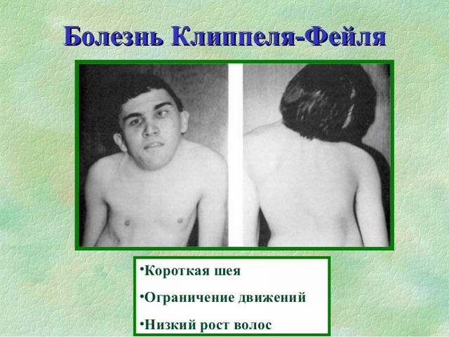
Essentially, short neck syndrome is a defect in the development of the cervical vertebrae. A visual manifestation of the disease is a sedentary and shortened, deformed neck. The pathology is characterized by the number of cervical vertebrae being less than normal, their small size compared to correctly developed vertebrae, and the fusion of the vertebral bodies.
Types of disease
In the practice of specialists, Klippel-Feil disease is quite rare. However, it is customary to distinguish several types of it:
- With a decrease in the physiologically correct number of cervical segments, they also grow together, which visually shortens the neck area even more. This form further complicates motor activity in the spine.
- Formation of synostosis of cervical structures - by their fusion with the structures of the skull. A person completely loses the ability to turn his head - due to a monolithic conglomerate formed by the vertebrae and occipital bone.
- The third type is a combination of the first two options. There may be pronounced synostosis of structures in the lumbar and lower thoracic regions.
Manifestation of pathology
Modern diagnostic methods allow a reliable diagnosis.
Klippel-Feil anomaly can manifest itself in a patient in three forms, which include:
- Reducing the number of segments in the cervical spine, which grow together and visually significantly shorten the neck. This form also causes difficult head movements.
- Cervical synostosis by fusion with the occipital bone. In this case, the patient cannot turn his head, since now his cervical vertebrae and the back of the head are one monolithic formation.
- The third form includes the first two, where synostosis of the lumbar and lower thoracic segments can also be observed.
The reasons for the formation of a certain form in a patient are not precisely explained.
Children's doctor
- Are you a medical student? Intern? Children's doctor? Add our site to social networks!
The syndrome, described in 1912 by Klippel and Feil, is characterized by the fusion of the last cervical vertebrae with the first thoracic vertebrae, and many other anomalies in the development of the skeletal and nervous system.
Etiopathogenesis of Klippel-Feil syndrome.
The etiology is unknown. Klippel-Feil syndrome is congenital (always evident from birth) and familial in nature.
It is believed that we are talking about a violation of very early segmentation during intrauterine life, which causes the appearance of all anomalies, since cervical somites are already obvious by the end of the 4th week of conception.
Klippel-Feil syndrome is described in the medical literature under many other synonyms:
- syndrome of numerical reduction of cervical vertebrae;
- congenital fusion of the cervical vertebra;
- synostosis of the cervical spinal column;
- congenital short neck.
Symptomatology of Klippel-Feil syndrome.
Manifestations in the skeletal system:
- synostosis of the cervical vertebrae is noted at birth and is a constant and characteristic sign, with the help of which a reliable diagnosis is made. The number of cervical vertebrae is reduced due to fusion with the thoracic ones. Thus, patients have a very short neck with reduced mobility (giving the patient the appearance of a “man without a neck” or with a “fixed neck”);
- kyphosis, cervical sclerosis or corticalis caused by synostosis of the cervical vertebrae.
Can be inconsistently combined:
- cervical spina bifida with or without meningocele;
- craniofacial asymmetry;
- defects of the ribs and fingers;
- cleft palate;
- the shoulder blades are located high.
- The most common pain is starting from the cervical and thoracic spine, spreading to the upper limb. They are caused by compression on the nerve roots or even directly on the spinal cord by protrusion of the intervertebral cartilage or an increase in the size of the vertebral body.
- External oculomotor nerve palsy.
- Cerebellar manifestations (equilibrium imbalance, nystagmus).
- Spastic hemiplegia.
- Hydrocephalus.
- Cramps.
- Hearing disorders (neurogenic origin).
Other associated clinical signs:
- low-set hair on the head (they go down under the biacromial line);
- hypoplasia of the trapezius or pectoral muscles;
- expressionless face;
- delayed mental development (more apparent than real).
Diagnosis of Klippel-Feil syndrome.
X-ray examination of the skeletal system facilitates the establishment of fusion between the cervical and thoracic vertebrae or reveals the presence of hemivertebrae.
Pathological examination reveals:
- complete or partial fusion of two or more cervical and thoracic vertebrae;
- deformation of the vertebrae or only the presence of hemivertebrae;
- small intervertebral foramina;
- the presence of some medullary tumors (angiolipomas);
- accessory lobes of the lung;
- non-closure of the foramen ovale;
- cystic formations along the length of the digestive tract.
Based on Klippel-Feil syndrome, Wildervanck in 1960 describes a more complex syndrome characterized by a clinical combination of anomalies of several syndromes:
- Klippel-Feil syndrome;
- Turk-Stilling-Daune syndrome (limited abduction of the eyeball; enophthalmia and binocular vision);
- labyrinthine deafness or deaf-muteness.
The syndrome described by Wildervanck is a very rare exception in medical practice (before 1970, only 20 cases were known, of which 19 patients were female).
The course and prognosis of Klippel-Feil syndrome.
READ ALSO: Prisoner syndrome
The course and prognosis are relatively favorable. All existing anomalies have little effect on the functional ability of the devices and systems of patients and thus - with little difficulty - they can be included in public life.
Treatment of Klippel-Feil syndrome.
There is no etiopathogenetic treatment.
Symptomatic treatment is always prescribed if the patient has clinical signs that cause him suffering.
Complications and prognosis for the patient
The disease is dangerous primarily due to its complications - renal failure, disruption of the functioning of the cardiovascular system and respiratory organs.
In addition, there is a decrease in sensitivity and atrophy of the muscle tissues of the upper and lower extremities.
Particularly severe situations can lead to complete immobilization of the neck, head and arms.
The prognosis for Klippel-Feil syndrome is quite favorable. The earlier the disease is detected, the better the effect of the treatment. Patients are able to lead a full life, albeit with some limitations.
After surgery and a rehabilitation period, the patient can lead a full life. During this time, the neck will increase slightly in length, which, from an aesthetic point of view, is considered a favorable aspect.
The latter are caused by constant pinching of the nerve roots and can lead to complete immobilization of the limbs. Damage to internal organs is dangerous due to irreversible pathological processes and can cause early death.
After surgery and recovery, the patient can move his head freely and lead a normal life. His neck will increase slightly in length, which is a favorable aspect from an aesthetic point of view.
In the case of a non-serious attitude, the patient not only develops diseases of the internal organs, but also experiences severe pain due to the formation of a pinched nerve root, which in severe cases can lead to complete immobility of the neck and limbs.
During the process of degenerative changes, numbness of the limbs and muscle atrophy can be observed.
According to experts, vertebral fusion does not pose any danger other than an aesthetic inconvenience.
Attention should be paid to complications, because they can significantly shorten a person’s life expectancy, provoke irreversible processes in diseases of internal organs, and also lead to the formation of pathologies of the respiratory system and heart disease.

Scoliosis often develops in Klippel-Feil syndrome
Due to a severe form of spinal underdevelopment and when parents refuse regular treatment of the child, complications from the nervous system develop. During growth, degenerative processes progress in the spinal column, which leads to deformation of the intervertebral discs.

Compression of the nerve roots causes disturbances in the sensory and motor spheres below the site of the lesion. Paresthesia appears in the arms and legs (a feeling of numbness and tingling), weakness of the muscles of the upper and lower extremities, and a decrease in temperature and pain.
Such pathological changes are typical for older children. In younger children, complications from the nervous system usually manifest themselves in the form of synkinesis - involuntary movements of the hands and arms.
Klippel-Feil syndrome: symptoms and treatment
Most doctors adhere to conservative treatment tactics for the disease. Patients suffering from this pathology are required to attend massage sessions and physical therapy. In case of pain, antispasmodic drugs and physiotherapy are prescribed.
In some cases, surgical treatment with resection of the periosteum is possible. The intervention allows for partial elimination of cosmetic defects and helps to increase the range of movements.
After surgery, the cervical spine is immobilized for the required period of time. The intervention is initially carried out unilaterally. As soon as the patient’s recovery has begun, doctors begin work on the opposite.
Unfortunately, modern medicine cannot offer adequate methods to completely eliminate Klippel-Feil syndrome. Treatment involves preventing the development of secondary deformities. Conservative methods are taken as a basis, which include exercise therapy and massage.
Drug treatment is prescribed if the pathological process is accompanied by pain and compression of the nerve roots. Typically, analgesics, anti-inflammatory and non-steroidal drugs are used.
Persistent pain caused by compression of the nerve roots is the main indication for surgery. In patients diagnosed with Klippel-Feil syndrome, the prognosis worsens with age. Therefore, surgery is prescribed immediately after confirmation of the pathology.

Bonola cervicalization is used to increase neck mobility. During the procedure, the doctor removes the four upper ribs and the periosteum to relieve pressure on the internal organs. The operation is carried out in several stages. Initially, the ribs are removed on one side, and after the body is restored, on the other.
The rehabilitation period requires a lot of time and effort. The patient must remain motionless for several months. However, you should not refuse the operation. Currently, this is the only effective way to correct external defects, the occurrence of which is provoked by Klippel-Feil syndrome. What health consequences may occur if you refuse treatment are described below.
Treatment tactics for Klippel-Feil syndrome must be comprehensive. The main emphasis is on activities such as massage courses, exercise therapy classes, and various physical procedures. They best help improve tone in the muscle groups of the back, improve local and general blood flow, as well as metabolism in the area of pathology.
READ ALSO: Symptoms and treatment of lumbar radicular syndrome
If persistent pain impulses appear and there is no effect from taking modern anti-inflammatory drugs and analgesics, surgical intervention is resorted to. In most cases, it is quite enough to carry out a cosmetic operation - to restore the physiological location of the muscle groups of the body in relation to the area of the shoulder blades.
The first step is to contact a competent specialist
Particularly severe variants of pathology require radical therapeutic manipulations, for example, modeling of spinal structures using modern implants.

The prognosis largely depends on the severity of pathological changes and the presence of complications from internal organs. It is especially unfavorable in the formation of degenerative processes in vertebral structures.
- Vertebro-basilar insufficiency against the background of cervical osteochondrosis
- Ointments for cervical osteochondrosis
- Pain in the neck
Related Posts
Klippel-Feil syndrome: developmental features and other defects
Klippel-Feil syndrome is a genetically determined disease. Pathological changes in the body begin to occur in the first weeks from the moment of conception. Among the main causes of the syndrome, doctors note a violation of the development and segmentation of the spine, especially at the upper cervical level.
Formed synostoses of the thoracic and cervical vertebrae, a decrease in their number, non-fusion of the bodies and arches make it possible to determine the clinical picture of the disease. The risk group includes children with unfavorable heredity, where the following is observed:
- Genetic defect in chromosomes. In a sick child, the formation of growth differentiation necessary for the full development of the skeleton occurs. This inevitably affects the development of the thoracic and cervical vertebrae.
- Autosomal dominant type of inheritance. If one of the parents has a disease, the probability of having a child with pathologies is 50-100%.
- Autosomal recessive type of inheritance. In this case, the probability of having a child with pathologies is 0-50%.
To avoid an unfortunate legacy, parents should consult a geneticist at the stage of planning a child.
One of the congenital anomalies of the structure of the spine is Klippel-Feil syndrome or short neck syndrome. According to ICD-10, it has the code Q76.1. This is a malformation of the vertebrae of the cervical and upper thoracic level, leading to shortening and deformation of the neck. It can be combined with other malformations of the musculoskeletal system.
The condition is congenital and begins in utero at the earliest stages of development - already in the first weeks after conception. If the changes in the vertebrae are not severe, then the syndrome may not be diagnosed immediately, but as the child grows or even after growing up.
The causes of Klippel-Feil syndrome are disturbances in the embryo of the processes of formation, fusion, development, and segmentation of the spinal column, especially at the upper cervical level.
As a result, the vertebral bodies are small, deformed, and fused with each other.
Embryogenesis disorders are caused by an anomaly in the 5, 8, 12 pairs of chromosomes, which leads to a disruption in the production of growth differentiation factor. It is responsible for the formation of the boundary between bone and cartilage tissues. Autosomal dominant and autosomal recessive inheritance are possible, which is less common.
Classification
There are 3 types of Klippel-Feil syndrome. Distribution among them is made on the basis of X-ray data.
- Type 1 – characterized by a decrease in the number of cervical vertebrae;
- type 2 – a single monolithic formation is formed, including the occipital bone, the cervical part of the spine and the upper thoracic vertebrae;
- Type 3 – not only the cervical, but also the lower thoracic and lumbar vertebrae are pathologically changed.
The syndrome can be combined with an anomaly of the scapula. In this case, they have a high location and irregular osteochondral structure (synchondrosis). In this case, a diagnosis of Kleppel-Feil-Sprengel disease is made.
In addition, there are often other developmental defects of the musculoskeletal system and internal organs. Thus, an increase in the number of fingers (polydactyly), their underdevelopment or fusion (syndactyly), pterygoid cervical folds, anomalies of the maxillofacial region, pathological forms of the skull, scoliosis, spina bifida (spina bifida or spina bifida) are possible.
Anomalies in the structure of the kidneys and ureters, the heart and large vessels, and the central nervous system are revealed. If the brain is damaged, there may be epileptic seizures and mental retardation.
If other developmental defects are identified, they are indicated additionally in the diagnosis.
Kleppel-Feil syndrome has 3 mandatory external signs:
- low hair growth line at the back of the head,
- shortened neck
- decreased range of motion.
Definition
Sliding hernias are those hernias in which, in addition to the parietal sac, the formation of the hernial sac also involves the visceral peritoneum, covering a certain segment of the sliding organ, and its other parts are devoid of a serous membrane, located in the retroperitoneal, extrahernial sac, in the retroperitoneal or preperitoneal tissue.
Sliding inguinal hernias are observed in older people, often in men. If the organ is located mesoperitoneally, exits through the internal ring of the inguinal canal with that wall that is not covered by the peritoneum, then the protrusion does not have a hernial sac.
Causes
SG is a type of abdominal hernia; their occurrence is caused mainly by the same factors as the occurrence of ordinary hernias.
The causes of hernia formation are divided into local and general. Local causes lie in the peculiarities of the anatomical structure of the inguinal or other area where the hernia occurs, and therefore are considered when studying individual types of hernias.
The common causes of hernia formation are divided into those that contribute and those that cause. The first include: heredity, age, gender, physique, weight loss, changes in internal organs and profession.
Heredity occurs in approximately 20 - 25%. Most often, hernias occur in children in the first year of life. At 10-20 years of age, their frequency increases again, reaching a maximum peak at 30-50 years.
One of the main causes of hernias in children is non-fusion of the peritoneal process. The process of fusion of the peritoneal process is associated with the process of descent of the testicle into the scrotum. Fusion of the peritoneal process occurs because there are ring-shaped fibers in the human cremaster. Contracting, these fibers compress the serous tube, it gathers into folds that grow together and gradually the peritoneal process turns into a connective tissue cord. These ring-shaped fibers are concentrated, like sphincters, in three places: the first sphincter at the top, at the level of the site of formation of the cremaster, the second in the middle part of the spermatic cord, and the third at the bottom of the testicle. It is in these places that the process of fusion of the peritoneal process begins, and it can heal completely or partially, remaining open at the top, on the side of the abdominal cavity, or at the bottom on the side of the testicle, or somewhere along the spermatic cord. The process of fusion sometimes ends only in the first months of a newborn’s life. The peritoneal process may not close at all and remain open from the abdominal cavity to the testicle; this is a consequence of underdevelopment of the cremaster.
In a female fetus, the peritoneal-inguinal process in most cases heals already at the beginning of its development, which explains the absence of inguinal hernias in girls.
The predominance in the number of oblique inguinal hernias over straight ones in children is explained by the fact that in newborns the large bladder closes the supravesical cavities of the anterior abdominal wall, leaving free only the lateral fossae, which correspond to the internal opening of the inguinal canal. Even a slight increase in intra-abdominal pressure promotes the entry of the viscera into the peritoneal-inguinal process.
The occurrence of hernias depends on the structure of the body and muscles and the shape of the abdomen. The occurrence of hernias is most facilitated by the female shape of the abdomen, followed by a cylindrical or mixed shape. Persons with a female abdomen are susceptible to inguinal hernias.
In persons with a male abdomen, the inguinal rings are stable even with significant physical exertion.
Among the general factors that cause the formation of hernias, those that increase intra-abdominal pressure should be put in first place, and those that weaken the abdominal wall in second place.
The first include: 1) disorders of the act of defecation - diarrhea, 2) cough 3) screaming, 4) difficulty urinating 5) tight girdling (bandaging) of the abdomen; 6) singing, playing wind instruments; 7) childbirth, 8) hard work, etc.
Factors that weaken the abdominal wall include: 1) pregnancy 2) old age, 3) a- and physical inactivity 4) general diseases that reduce muscle strength 5) damage to the abdominal wall.
Symptoms
There are no clearly pathognomonic symptoms of HS hernias. The patients complained of constant dull pain in the hernia. In the sigmoid colon, pain radiates to the sacrum, or to the rectum, to the anus. There is increased pain in the hernia when straining and sitting.
Constipation and bloating occur in patients with ordinary abdominal hernias and therefore do not have significant diagnostic value for recognizing FH of the colon.
One of the typical signs of HS of the colon is incomplete reducibility of the hernia. Unreduced hernias are usually divided into hernias with complete and partial irreducibility. Partial (incomplete) reducibility is a characteristic feature of congenital HS of the cecum. The medial part of the hernial contents is completely reduced, but the lateral part remains unreduced.
Trevs's symptom is explained by the fact that, together with the cecum, loops of the small intestine enter the hernial sac, which easily move into the abdominal cavity when pressing with the hand on the hernial rupture and the cecum, which is an integral part of the wall of the hernial sac and fixed from the outside of the hernial orifice, is not reduced. The described anatomical relationships explain the relative constancy of the size of the SG in general and the large intestine in particular. This symptom is also one of the characteristic symptoms of FH of the colon. It manifests itself in the fact that after reduction of the hernial indentation, it immediately comes out again, regardless of the vertical or horizontal position of the patient.
A hernial bulge in HS is palpated as a soft, pasty formation, caused by the presence of a colon wall devoid of a serous membrane.
The patient complains of constant pain in the hernia, which intensifies with tension.
There are a number of symptoms based on which a bladder hernia can be diagnosed in time. Patients with SG of the bladder often complain of urinary disorders, in particular urinary retention or frequency, sometimes the so-called. double act urination.
The clinical picture of strangulation of the internal female genital organs resembles the clinic of intestinal strangulation. However, strangulation of the ovary leads to sharp pain in the hernia in the lower abdomen.
Classification
A classification is given, including 4 types of sliding inguinal hernias of the colon:
- Intraperitoneal SG, when the colon is covered on all sides by the peritoneum, except for the place of its attachment to the hernial sac. Unlike a regular hernia, the intestine is not freely located in the hernial sac, but is fused with it by its mesentery. In essence, we are talking about the sliding of the mesentery of the intestine attached to the pouch, and not the intestine itself. Separation of the hernial sac from the intestine during surgery is associated with the risk of damage to its vessels.
- Paraperitoneal SG, when the intestine is partially retroperitoneal, forming one of the walls of the hernial sac. It is covered with a serous membrane on one side - the retroperitoneal position of the intestine, or on three sides - its mesoperitoneal location.
- Intraparaperitoneal HS is an intermediate type and has elements of the previous two. In the aboral part, located intraperitoneally, the slipped intestine is fused with the hernial sac by the mesentery, and in the oral part the intestine is located paraperitoneally.
- Extraperitoneal SG, when the intestine lies retroperitoneal and the hernial sac is absent.
Diagnostics
 Diagnosis of FH is difficult and sometimes impossible. Preoperative diagnosis of sliding hernias is perhaps of utmost importance, as it allows you to draw up a surgical plan in advance and avoid damage to the organ that has slipped into the hernia.
Signs and methods for diagnosing HS depend on the complexity and the organ that has slipped into the hernia.
Mostly constant pain in the area of the hernial protrusion, which intensifies with walking and straining. Elderly and senile patients; Mostly men are affected. A large hernia, almost always with a wide hernial orifice, a moderate, wide and “soft” opening of the inguinal canal; the belly has a saggy shape.
Partial or complete irreducibility of the hernia (Trevs's symptom); sometimes the hernial protrusion consists of two parts: on the medial side it is reduced, but on the lateral side it is not reduced ("double" Trevs's hernia). A constant volume of the hernia with changes in the patient’s position (vertical, horizontal), tension, coughing, etc.. A certain pastiness of the hernia, sometimes it is possible to palpate a thickening of the intestinal wall; sometimes there is a vermiform appendix and appendices epiploicae. Tympanitis in the area of the hernia, which disappears when the colon is filled with fluid per rectum;
X-ray examination with a contrast suspension of the colon per os, or better yet per rectum, sometimes makes it possible to diagnose a hernia of the colon. Valuable methods for diagnosing SG of the bladder are cystoscopy and cystography. In the presence of dysuric phenomena and large hernias, endoscopy, radiocontrast cystography or pneumatoradiography of the bladder are performed.
Prevention
Hernias cause patients a number of sufferings, limit their ability to work, and in many cases lead to serious complications. There is no doubt about the need for early surgical treatment of patients for hernias.
This also applies to patients with HS, in whom complications occur more often than in patients with ordinary abdominal hernias. Such complications include, first of all, pinching; in addition, with HS, adhesions are formed that disrupt the anatomical relationship of the contents of the hernia and the hernial sac, increasing the risk of damage to organs and other anatomical structures during surgery. Therefore, the sooner surgery is performed, the lower the risk for the patient.
Inflammation of the hernia occurs due to infection of the hernial sac ( inflammation from within ) or hernia membranes ( inflammation outside ).
In case of infection from the abdominal cavity:
the general condition of patients quickly deteriorates,
signs of peritonitis progress,
intoxication increases.
The hernia increases in size as a result of edema and tissue infiltration,
subsequently skin hyperemia appears.
Hernia membranes can become inflamed as a result of primary damage to the skin (boils, abrasions, scratching). In such cases, the general condition of the patients suffers slightly.
Treatment
With the help of emergency surgery, the source of peritonitis and infection of the hernial sac are eliminated. In such cases, hernial orifice repair is not performed. In case of external infection, hernia repair can be performed only after the elimination of inflammatory processes in the skin.
Forecast
Prognosis for external abdominal hernias depends on the timeliness of the operation. Planned hernia repair is practically not accompanied by mortality associated with surgical intervention.
If complications develop, Firstly strangulated hernia, the danger to the patient’s life increases as the the period of time elapsed from the moment of injury to the operation.
Mortality after operations performed in the first 6 hours from the moment of infringement, is 1.0-1.5%.
In cases strangulated bowel resection mortality increases to 10-15%,
A with phlegmon of the hernial sac exceeds 25%.
Hernia of the white line of the abdomen
The linea alba runs from the xiphoid process to the symphysis pubis. From the point of view of surgical practice, it is important area between the xiphoid process and the umbilicus, where the width of this anatomical formation reaches 2 cm .
Hernias of the white line of the abdomen are more common epigastric and paraumbilical.
Hypogastric hernia are extremely rare.
Linea alba
It is a connective tissue plate formed by obliquely directed tendon fibers intersecting along the midline. In this case, a kind of mesh is formed in which there are slit-like spaces of various sizes filled with fatty tissue.
The root cause of hernias becomes an expansion of the white line of the abdomen. In men, it is often of a constitutional nature and is a consequence of abdominal obesity (“ beer belly"), and in women - the result repeat pregnancies.
Preperitoneal fiber penetrates the gap between the tendon fibers, and the so-called “ preperitoneal lipoma", capable of being infringed.
Subsequently, a hole in the white line of the abdomen under the influence of ysh The established intra-abdominal pressure gradually increases, a defect is formed into which the parietal layer of the peritoneum prolapses.
The contents of the hernial sac most often become stuffing box.
Epigastric hernias rarely exceed in diameter 4 -5 cm, in obese patients are not always visible and may be multiple.
To detect a hernia It is convenient to carry out palpation with the patient lying in a horizontal position, raised on his elbows.
A hernia of the linea alba can be accompanied by various gastrointestinal diseases, so differential diagnosis is necessary, including the use of the necessary instrumental research methods.
Inguinal hernia Kuzin
Inguinal hernias account for 75-80% of the total number of hernias. They are diagnosed mainly in men (90%). This is due to the fact that the inguinal canal in women is narrower and longer than in men, has a slit-like shape, and is better strengthened by muscle and tendon layers. In pediatric surgery, inguinal hernias are encountered even more often; they account for 95% of all ventral hernias. The main varieties are represented by oblique and direct inguinal hernias. There are also oblique hernias with a straight canal, preperitoneal, intramural, peri-inguinal, supravesical, etc.
Embryological information. From the third month of intrauterine development of the male embryo, the process of descent of the testicles begins. In the area of the internal inguinal ring, a protrusion of the parietal peritoneum forms, and processus vaginalis. In subsequent months of intrauterine development, further protrusion of the peritoneal diverticulum into the inguinal canal occurs. By the end of the seventh month, the testicles begin to descend into the scrotum. By the time the child is born, the testicles are in the scrotum, and the processus vaginalis of the peritoneum is overgrown.
When it fails to heal, it forms congenital inguinal hernia.
In case of incomplete infection of the vaginal process of the peritoneum, hydrocele of the spermatic cord occurs in certain areas of it ( funicolocele).
Anatomy of the groin area .
When examining the anterior abdominal wall from the inside from the abdominal cavity (Fig. 10.3), you can see five folds of the peritoneum and depressions (pits), which are the places where hernias emerge. The external inguinal fossa is the internal opening of the inguinal canal; it is projected approximately above the middle of the inguinal (pupart) ligament, 1 - 1.5 cm above it. Normally, the inguinal canal is a slit-like space filled in men with the spermatic cord, in women with the round ligament of the uterus. The inguinal canal runs obliquely at an angle to the inguinal ligament and ends at the external opening. In men, it has a length of 4-4.5 cm. The walls of the inguinal canal are formed: the anterior one - by the aponeurosis of the external oblique abdominal muscle, the lower one - by the inguinal ligament, the posterior one - by the transverse abdominal fascia, the upper one - by the free edges of the internal oblique and transverse abdominal muscles.
The external (superficial) opening of the inguinal canal is formed by the legs of the aponeurosis of the external oblique abdominal muscle, one of them is attached to the pubic tubercle, the other to the pubic fusion. The size of the external opening of the inguinal canal varies. Its transverse diameter is 1.2-3 cm. In women, the external opening of the inguinal canal is slightly smaller than in men.
The internal oblique and transverse abdominal muscles, located in the groove of the inguinal ligament, approach the spermatic cord and are thrown over it, forming the inguinal space of different shapes and sizes. The boundaries of the inguinal space are: below - the inguinal ligament, above - the edges of the internal oblique and transverse abdominal muscles, on the medial side - the outer edge of the rectus abdominis muscle. The inguinal space can have a slit-like, fusiform or triangular shape. The triangular shape of the inguinal space indicates weakness in the groin area.
At the site of the internal opening of the inguinal canal, the transverse fascia bends funnel-shaped and passes onto the spermatic cord, forming a common vaginal membrane of the spermatic cord and testicle.
The round ligament of the uterus at the level of the external opening of the inguinal canal is divided into fibers, some of which end on the pubic bone, the other is lost in the subcutaneous fatty tissue of the pubic region.
Congenital inguinal hernia. If the vaginal process of the peritoneum remains completely unfused, then its cavity freely communicates with the peritoneal cavity. Subsequently, a congenital inguinal hernia is formed, in which the vaginal process becomes a hernial sac. Congenital inguinal hernias make up the bulk of hernias in children (90%), but they also occur in adults (about 10-12%).
Acquired inguinal hernia.
The main reason for the formation of inguinal hernias is weakness of the posterior wall of the inguinal canal.
Distinguish indirect inguinal hernia and direct. An oblique inguinal hernia passes through the external inguinal fossa, a direct one - through the internal one.
At channel form the bottom of the hernial sac reaches the external opening of the inguinal canal.
At cord form the hernia exits through the external opening of the inguinal canal and is located at different heights among the elements of the spermatic cord.
At inguinal-scrotal form the hernia descends into the scrotum, stretching it.
Indirect inguinal hernia has an oblique direction only in the initial stages of the disease. As the hernia enlarges, the internal opening of the inguinal canal expands in the medial direction, pushing the epigastric vessels inward. The more medially the hernial orifice expands, the weaker the posterior wall of the inguinal canal becomes. With long-standing inguinal-scrotal hernias, the inguinal canal acquires a straight direction, and its superficial opening is almost at the same level as the internal opening ( indirect hernia with straightened course). With large hernias, the scrotum increases significantly in size, the penis is hidden under the skin, and the contents of the hernia do not move into the abdominal cavity on their own. When repositioning, a rumbling sound is heard in the intestines.
Direct inguinal hernia exits the abdominal cavity through the medial fossa, protruding the transverse fascia (posterior wall of the inguinal canal). Having passed through the external opening of the inguinal canal, it is located at the root of the scrotum above the inguinal ligament in the form of a rounded formation. The transversalis fascia prevents the direct inguinal hernia from descending into the scrotum. Often direct inguinal hernia is bilateral.
Sliding inguinal hernia are formed when one of the walls of the hernial sac is an organ partially covered by the peritoneum, for example the bladder, cecum and ascending colon (Fig. 10.4). Rarely, the hernial sac is absent, and the entire protrusion is formed only by those segments of the slipped organ that are not covered by the peritoneum.
Sliding hernias account for 1 - 1.5% of all inguinal hernias. They arise as a result of mechanical contraction by the peritoneum of the hernial sac of the adjacent segments of the intestine or bladder, devoid of serous cover.
Clinical picture and diagnosis.
It is not difficult to recognize a formed inguinal hernia. Typical is anamnesis:
sudden onset of a hernia during physical exertion
or gradual development of hernial protrusion
the appearance of protrusion when straining in a vertical position of the patient’s body and
reduction - horizontal.
Patients are concerned about pain in the hernia area, in the abdomen, and a feeling of discomfort when walking.
Inspection the patient in an upright position gives an idea of the asymmetry of the groin areas.
If there is a protrusion of the abdominal wall, its size and shape can be determined.
Finger examination The external opening of the inguinal canal is performed in a horizontal position of the patient after reduction of the contents of the hernial sac.
The doctor, using his index finger, the palmar surface of which is facing the posterior wall of the inguinal canal, in order to determine the condition of the posterior wall, invaginating the skin of the scrotum, enters the superficial opening of the inguinal canal, located medially and slightly higher from the pubic tubercle.
Normally, the superficial opening of the inguinal canal in men allows the tip of the finger to pass through. When the posterior wall of the inguinal canal is weakened, it is possible to freely place the tip of the finger behind the horizontal branch of the pubic bone, which cannot be done with a well-defined posterior wall formed by the transverse fascia of the abdomen.
Determine the symptom of cough impulse.
Both inguinal canals are examined.
Examination of the scrotal organs (palpation of the spermatic cords, testicles and epididymis) is mandatory.
Diagnosis of inguinal hernias in women is based on inspection and palpation, since it is almost impossible to insert a finger into the external opening of the inguinal canal. In women, an inguinal hernia is differentiated from a cyst of the round ligament of the uterus located in the inguinal canal. Unlike a hernia, it does not change its size when the patient is in a horizontal position; the percussion sound above it is always dull, and tympanitis is possible above the hernia.
Indirect inguinal hernia unlike direct more common in childhood and middle age;
it usually descends into the scrotum and sometimes bilateral.
With an indirect inguinal hernia, the posterior wall of the inguinal canal is well defined,
The direction of the cough impulse is felt from the side of the deep opening of the inguinal canal.
The hernial sac passes through the elements of the spermatic cord, therefore, upon objective examination, thickening of the spermatic cord on the side of the hernia is noted.
Direct inguinal hernia happens more often in older people.
The hernial protrusion is round in shape, located at the medial part of the inguinal ligament.
The hernia rarely descends into the scrotum, usually bilateral;
During an objective examination, the posterior wall of the inguinal canal is always weakened.
The cough impulse is felt directly against the external opening of the inguinal canal.
The hernial sac is located medially from the spermatic cord.
Sliding inguinal hernia has no pathognomonic signs. Usually this is a large hernia with a wide hernial orifice. Occurs mainly in individuals elderly or senile age. Diagnosis of sliding hernias of the colon is complemented by irrigoscopy.
With sliding hernias of the bladder, the patient may notice urination disorders or urination in two stages: first the bladder is emptied, and then, after pressing on the hernial protrusion, a new urge to urinate appears and the patient begins to urinate again. If a sliding hernia of the bladder is suspected, it is necessary to perform catheterization and cystography. The latter can reveal the shape and size of the bladder hernia and the presence of stones in it.
Differential diagnosis.
An inguinal hernia should be differentiated from a hydrocele, varicocele, and also from a femoral hernia. An erroneous diagnosis of an inguinal hernia can be made when inguinal cryptorchidism (both unilateral and bilateral), when there is a delay in the descent of the testicle into the scrotum. Such patients are bothered by pain and swelling in the groin area. The correct diagnosis is easy to make after palpation of the scrotum. However, it must be remembered that 25% of patients with cryptorchidism have a concomitant inguinal hernia. Untreated inguinal cryptorchidism is often complicated by the development of seminoma, a rapidly growing malignant testicular tumor, since the testicle located in the inguinal canal is much more often injured and overheated than a normally located one.
An irreducible inguinal hernia, causing an enlargement of the scrotum, becomes similar to hydrocele (dropsy of the testicular membranes). In this case, fluid accumulates between the layers of the testicular membrane and, as a result, the size of the scrotum increases. The difference between a hydrocele and an irreducible inguinal-scrotal hernia is that it has a round or oval rather than pear-shaped shape, a dense elastic consistency, and a smooth surface. The palpable formation cannot be distinguished from the testicle and its epididymis. A large hydrocele, reaching the external opening of the inguinal canal, can be clearly separated from it by palpation. The percussion sound above the hydrocele is dull; above the hernia it may be tympanic.
An important method of differential diagnosis is diaphanoscopy (transillumination). It is performed in a dark room using a flashlight held tightly to the surface of the scrotum. If the palpable formation contains clear liquid, then it will have a reddish color when examined. The intestinal loops and omentum located in the hernial sac do not allow light rays to pass through.
Similar to inguinal hernia varicocele(varicose veins of the spermatic cord), in which when the patient is in an upright position, dull arching pain appears in the scrotum and a slight increase in its size is noted. Upon palpation, you can detect a serpentine dilatation of the veins of the spermatic cord. Dilated veins easily collapse when pressure is applied to them or when the scrotum is lifted upward. It should be borne in mind that varicocele can occur when the testicular vein is compressed by a tumor of the lower pole of the kidney.
Treatment. The main method is surgical treatment. The main purpose of the operation is plastic surgery of the inguinal canal.