Causes of acquired cataracts in infants. Causes, symptoms, methods of treating congenital and other types of cataracts in children. Infectious and metabolic pathologies in the genesis of cataracts
Cataracts in children can appear at any age. In children, a deviation in which the lens becomes cloudy does not cause any pain or discomfort, which is especially dangerous. Parents are often unaware of the problem, and developing cataracts in a child can lead, if not to loss of vision, then to its deterioration. More often, the pathology is observed in the form of a congenital anomaly. Changes in the eyeball are usually eliminated surgically. If the clouding of the lens is localized in the periphery and central vision is not affected, no intervention is required. Acquired cataracts are detected less frequently in children.
It is not always possible to determine what factors contributed to the development of cataracts. The reasons are different. Congenital pathology in most cases is hereditary.
The occurrence of abnormalities in the baby's eyeball could be provoked by infectious diseases of the mother during pregnancy in the form of influenza, measles, rubella, syphilis, tocoplasmosis, as well as the presence of herpes and cytomegalovirus in her body.
Congenital cataracts sometimes appear in premature babies. The cause of the abnormality in the baby may be the use of antibiotics by a pregnant woman. Often children are born with pathology whose mothers have diabetes.
The development of deviation in newborns is associated with internal factors. Acquired cataracts in a child occur due to external causes such as:
- increased radiation;
- bad environment;
- mechanical injury;
- unsuccessful operation;
- infectious disease;
- inflammatory process;
- metabolic problems;
Abnormal development of the eyeball can be triggered by brain injury, prolonged use of medications, the presence of toxacariasis, or a failure in the production of galactose.
Features and types of congenital pathology
A newborn baby may have cataracts in the front of the lens. Its appearance is associated with a genetic factor. With this type of pathology, vision is not impaired. Treatment does not require surgical intervention.
A deviation in a child is sometimes detected in the back of the lens. Nuclear cataract is diagnosed at its center. It is rare in children. With a layered bilateral appearance, the visual pathology falls below 0.1, and opacification is detected around the nucleus. The danger comes from complete cataracts, since the anomaly affects all layers.
The fact that the baby has a deviation is indicated by a whitish or gray tint of the pupil.
He rubs his eyes with his hands, unable to focus on the toy. The child does not watch a moving object or looks at it with one eye. With congenital cataracts, you may notice opacities in the form of dots on the pupil or on both.
 Lack of timely intervention in such a disorder threatens the development of amblyopia or strabismus. And this often leads to problems at school in the future and affects the development of the child’s personality. If symptoms are observed that indicate a deviation, an examination by an ophthalmologist is necessary. Using special equipment and drops that dilate the pupil, the doctor will see the condition of the eyeball.
Experts believe that if central vision decreases due to clouding of the lens, then cataracts that arose at the birth of the baby must be removed no later than the date when the child turns 3 months old. However, there is a risk of increased intracranial pressure. At this age, it is difficult for a baby to endure anesthesia.
If there is a small area of opacity and there is no problem with central visual acuity, surgery is not required, but the condition of the eyeball is constantly monitored.
Symptoms of the disorder in older children
Developing cataracts prevent the baby from perceiving normally the world that surrounds him. Older children have even more problems. If there is a deviation, which is associated with clouding of the lens, they experience:
- Flashing dots in the eyes.
- Blurred look.
- Decreased visual acuity.
- Splitting of objects.
Acquired cataracts, like congenital ones, can develop in different ways. Each type of pathology has its own characteristics in treatment.
With the subcapsular form, which occurs in children with diabetes, the back surface of the lens is damaged, with the polar form - the entire area in both eyes. The development of secondary cataracts is facilitated by chronic illness or unsuccessful surgery. The traumatic type is preceded by exposure to chemicals. Bilateral damage to the lens with layered cataracts threatens complete loss of vision.
Sometimes the pupil does not take on a different color. The baby sees normally and does not turn to look at the object. Parents do not notice anything abnormal. And already at school, straining his eyes, the child will complain that a spot near the pupil is bothering him.
Point cataract, which is diagnosed quite rarely, occurs:
- due to intrauterine infection;
- in premature infants;
- with chromosomal disorders.
Diabetes and Wilson's disease can provoke changes in the lens. The cause of this type of cataract can be a decrease in calcium in the blood plasma, as well as hypoglycemia, in which the glucose level decreases. A child with such a deviation sometimes has not one, but several dots on the pupil. The protein takes on a milky hue. The cloudiness is localized in different parts of it. The baby often has the urge to rub his eye.
In adolescents, it provokes the development of cataracts:
- Smoking.
- Drinking alcohol.
- Unhealthy environmental situation.
Sun radiation and exposure to chemicals contribute to the disorder. It gets to the lens when the eye is injured.
Diagnosis and treatment

Before the procedures, a solution is instilled into the pupil, which helps to dilate it. Schoolchildren are checked for angle and visual acuity, the retina is examined, and the pressure inside the eye is measured.
Having identified the degree of clouding, the stage of cataract is diagnosed - from initial to overripe. The absence of deviation is indicated by a transparent lens. Pathology is not treated by resorting to folk recipes or physiotherapeutic procedures. Pharmaceutical drugs do not help with this disorder. In case of peripheral damage, monitor the condition of the eyeball.
The operation is performed when clouding of the lens spreads over its entire surface and vision deteriorates.
The type of intervention depends on the age of the baby; the general condition and the presence of other pathologies are taken into account. The lens can be removed together or without a capsule. Using cryoextraction, they freeze it and then remove it. During phacoemulsification, ultrasound is applied to the eye. Now special lenses are produced that can replace the affected element. They perform the function of a lens.
Ignoring such a disease in a child can lead to serious consequences in the form of:
- glaucoma;
- myopia;
- swelling of the eyeball;
- inflammatory process;
- blindness.
One of the most common eye diseases is cataracts. It is mainly diagnosed in adults and the elderly, but it can also be found in children.
The prevalence rate in newborns is 5 people per 100 thousand, in older children - 3-4 cases per 10 thousand people.
Definition of disease
Cataract is an eye disease in which clouding of the lens material occurs with partial or complete loss of visual acuity and clarity. Cloudiness can be either total or incomplete.
According to the International Classification of Diseases, 10th revision, nosology is encrypted as H25-H28. But congenital disease in children according to ICD-10 has the code Q12.0.
The lens is a biconvex lens that refracts the sun's rays passing through it and focuses them on the retina.
Stimulation from the retina is transmitted along the optic nerve to information processing areas in the brain.
With cataracts, due to clouding, the refraction of sunlight is disrupted, the image becomes blurry.
Etiology
It is not possible to find the exact cause of cataracts, but there are factors that may predispose to its development:
The leading factor in the appearance of congenital cataracts is heredity. Often among close relatives of a sick child (mother, father, brothers and sisters) cases of cataracts in the anamnesis are identified.
The disease is linked to certain genes, and there is a high probability of cataracts in the offspring.
Causes of congenital pathology in children:
But congenital cataracts are also registered in children without family history. How can this be explained?
The fetus is very susceptible to viral infections in the first trimester of pregnancy.
If at this time it is attacked by viruses, then the congenital form may develop and will become the least of the evils that viruses can cause to the fetus.
Pathogens of intrauterine infection:

In diabetes mellitus, there is an increase in glucose content in the lens due to hyperglycemia. The fibers of the lens swell and lose their transparency - this is how this type of cataract begins.
With galactosemia, galactose accumulates in the lens in a similar way. In transmitted light it looks like oil drops. These accumulations can be seen already during the first days of a child’s life.
For traumatic lesions, regardless of age rosette cataracts occur, which progress and can completely occupy the entire lens.
Cloudiness of the lens can occur as a complication of other diseases. For example, with uveitis, inflammatory products can penetrate into the lens, which leads to the development of cataracts.
Various radiations have a negative effect on the lens: infrared, ultraviolet. Peeling of the anterior chamber of the lens occurs, which leads to its clouding.
When there is a deficiency of calcium ions in the body, calcium cataracts occur. Its development is possible with the removal of the parathyroid glands, which are responsible for calcium metabolism.
Cloudiness appears in the form of small, sometimes bright spots on the pupil, which can be seen with the naked eye. Treatment of children with pinpoint cataracts is long-term.
Chronic use of certain drugs can also lead to illness. The list includes hormonal drugs and cardiac glycosides.
The ingress of various substances, such as alkalis, leads to toxic cataracts. Alkali reduces the acidity of the anterior chamber of the eye, glucose is washed out of the lens.
Causes, symptoms and treatment of the disease:
Classification
Depending on the age of onset of cataracts, there are 2 types of cataracts - congenital and acquired.
More often, ophthalmologists encounter acquired cataracts; congenital cataracts are quite rare.
Depending on the stage there are:
- initial;
- immature;
- mature;
- overripe.

Clinical manifestations
Cataracts are detected in a newborn child usually at periodic medical examinations - you should not avoid them. You can suspect cataracts in a child in the following cases:
- the child practically does not react to silent toys;
- does not follow the parents' gaze - does not focus vision;
- rapid uncontrolled eye movements;
- the pupil is gray or white.
- examination and assessment of condition;
- operation;
- rehabilitation.
- Congenital. It is either already present in the child at birth or appears within a few days after.
- Acquired. It may appear at least two months after birth.
- Genetics. Typically, congenital cataracts in children appear due to a “breakdown” in genes received from both parents. Next, the lens of the eye begins to develop incorrectly.
- Chromosomal abnormalities related to various pathologies. For example, Down syndrome.
- Infections suffered by the mother during gestation are toxoplasmosis (a helminth infection that can be contracted through water, soil, and also from pets); rubella (virus, the body becomes covered with a red blistering rash); chickenpox (a mild form of chickenpox); cytomegalovirus (a common viral disease that is often asymptomatic and has unpleasant consequences); herpes virus.
- Eye damage. For example, during an injury or after surgery.
- . With this pathology, the child’s body cannot break down galactose.
- Toxocariasis. A rare disease that affects the eyes. Toxocariasis is most often contracted from animals.
- one or both pupils of the child have become white or gray;
- the child is not able to focus, for example, on a toy, he inactively watches moving objects and people;
- The child's eye movements become uncontrollable.
- Dill seeds. Take 3 teaspoons of dill seeds, place them in 2 fabric bags (it should hold water well) measuring five by five centimeters. Pour water into the bags with seeds and boil. Then cool them and apply to your eyes. Keep the compress for 15 minutes.
- Calendula. Take 2 teaspoons of calendula officinalis flowers and pour 2 cups of boiling water. The resulting infusion can be used internally and can also be used to wash the eyes.
- Parsley and carrot juices. You need to combine them in a ratio of one to ten, mix well. Drink 3 times a day.
- Spinach and carrot juices. Combine in a ratio of one to five. Drink 3 times a day.
- Surgery using cryoextraction method. The lens is extracted by “sucking” it to the cold tip of the cryoextractor.
- Operation using the intracapsular extraction method. Using an ice-cold metal rod, the lens is removed along with the capsule. With extracapsular extraction, the capsule is not removed, but remains in the eye. The core and substance are removed.
- Phacoemulsification. During surgery using this method, several two to three millimeter incisions are made. Through them, the lens is exposed to ultrasonic radiation. It is converted into an emulsion, then removed. A special elastic lens is placed where the lens used to be. It takes root well inside the eye.
- Women planning a pregnancy need to cure chronic diseases or put them into stable remission.
- During pregnancy, you need to avoid exposure to radiation, toxic waste, and viral diseases.
- Children should be given vitamins for the eyes (B1, B2, B12 and others prescribed by a doctor). Nutrition should be balanced.
- After the operation, you need to support the child and follow the advice of ophthalmologists.
- chromosomal abnormalities: trisomy 21, Turner syndrome, trisomy 13, trisomy 18, cry-the-cat syndrome;
- craniofacial syndromes: cerebro-oculofacial skeletal syndrome, nephropathy, Lowe's syndrome, Allport's syndrome, Hallermann-Streiff-Francois syndrome;
- skeletopathies: Smith-Lemli-Opitz syndrome, Conradi syndrome, Weill-Marchesani syndrome, Stickler syndrome, Bardet-Biedl syndrome, Rubinstein-Taybi syndrome, syndactyly and polydactyly;
- neurometabolic diseases: Zellweger syndrome, Meckel-Gruber syndrome, Marinescu-Sjögren syndrome, infantile neuronal lipofuscinosis;
- myopathies: myotonic dystrophy;
- dermatological: crystalline cataract and uncombable hair, Cockayne syndrome, Rothmund-Thomson syndrome, atopic dermatitis, pigment incontinence syndrome, progeria, congenital ichthyosis, ectodermal dysplasia, Werner syndrome.
- intrauterine infection (, toxoplasmosis);
- infection acquired at an early age, e.g. Herpes Simplex(cataract as a consequence of previous uveitis or parsplanitis);
- medications (corticosteroids, chlorpromazine);
- metabolic disorders (galactosemia, galactokinase deficiency, hypocalcemia, hypoglycemia, diabetes mellitus, mannosidosis, hyperferritinemia);
- trauma (accident or intentionally caused damage, laser photocoagulation, radiation injuries);
- due to other diseases: endophthalmitis, retinopathy of prematurity, persistent primary vitreous, retinitis pigmentosa, aniridia, microphthalmia).
- appearing immediately after birth (congenital) or acquired over the course of life;
- hereditary and non-hereditary.
- fetal nuclear - opacification of the lens substance between the anterior and posterior Y-shaped sutures, which is often accompanied by opacification of the posterior capsule;
- cortical - opacification of the anterior or posterior cortical layers, not extending to the fetal nucleus and often accompanied by opacification of the posterior capsule;
- persistent fetal vasculature - the presence of one or more of the listed features (retrolental membrane with or without visible vessels, patent or impassable hyaloid vessel, distended ciliary processes);
- isolated opacification of the posterior capsule (posterior polar cataract) - opacification of the posterior capsule without opacification of the layers of the cortex lying closer to the center and the nucleus;
- posterior lentiglobus - posterior protrusion of the posterior capsule with or without a posterior capsule defect.
- partial;
- complete (total - the entire lens is diffusely cloudy).
- Unilateral - rarely accompanied by a systemic disease. A common cause of unilateral cataracts is persistent fetal vasculature. Another common cause of this type of cataract in children is trauma. It may develop after laser photocoagulation for retinopathy of premature infants or after intraocular surgery.
- Bilateral - the etiology is established in approximately 50% of children. The most common cause of this form in Europe and the USA is autosomal dominant hereditary cataract.
- For myopic refraction and astigmatism, the most complete correction is prescribed.
- For hypermetropia, the prescribed correction depends on the age of the child. Before one year of age, low-myopic refraction is formed with glasses.
- From two years of age and older, two pairs of glasses for distance and near, or one bifocal glasses, or complex progressive glasses are prescribed.
- direct - the better seeing eye is closed (prescribed for a period from several hours a day to a month, depending on the severity of amblyopia);
- alternately direct - the leading eye closes more often (the ratio of closing time is 1:2, 1:3, 1:6, and so on).
- flare (general flare according to Bangerter) - a large area of the retina is illuminated, its inhibition develops, and the area of the macula is the first to emerge from inhibition, since bright light is an adequate stimulus for the cones. After the exposure, the child is asked to perform a visual load at close range for 5-10 minutes;
- maculotester is a device for forming correct fixation;
- sensory training (computer pleoptics);
- physiotherapy (laser stimulation, magnetic stimulation).
- in the first year, inspection is carried out once every two months;
- in the second and third years - once every 3-4 months;
- subsequently - once every six months.
- Genetic predisposition is considered the most common cause of congenital cataracts.
- The birth of a baby ahead of schedule. Moreover, the earlier a child is born, the more immature his visual organs will be, and the higher the risk of developing cataracts.
- Diabetes mellitus in an expectant mother.
- Taking strong medications during pregnancy.
- Infectious and inflammatory diseases suffered by a woman during pregnancy.
- Negative effects of radiation.
- Brain damage.
- Traumatic eye injury.
- Uncontrolled use of medications.
- Infections, foci of inflammation.
- Metabolic disorder.
- Unfavorable environmental conditions.
- Smoking and drinking alcohol in adolescence.
- Negative effects of UV rays when the child does not wear sunglasses in sunny weather.
- Subcapsular posterior. Occurs due to damage to the posterior capsule of the lens. It is characterized by rapid progression, an increase in the size of the lens, and disruption of metabolic processes in the eye area. Treatment is surgical.
- Polar. Cloudiness occurs in the area of the anterior or posterior poles of the lens. With posterior localization, vision decreases slightly. The anterior form contributes to a significant decrease in vision, but is not prone to progression.
- Capsular. Cloudiness develops in the area of the anterior capsule, causing persistent and significant visual impairment.
- Layered. It is considered the most common. The affected layers of the lens alternate with healthy ones.
- Membranous. It occurs if the child had complete cataracts at birth, but the pathology has resolved for some reason. The lens is clouded by a specific film, which was formed from the remains of the clouded mass.
- Radial. Occurs as a result of the negative effects of UV rays.
- Traumatic. Develops with various types of traumatic lesions of the eyeball.
- Toxic. Occurs after taking medications.
- Exchange. It occurs as a result of diseases that disrupt metabolic processes in the body as a whole, and in the area of the eye whites, in particular.
- Complicated. Develops after suffering inflammatory eye diseases.
- Initial. At this stage, a watery infiltrate forms in the area of the optical lens.
- Immature. The affected area extends to the lens capsule, causing significant loss of visual acuity.
- Mature. Cloudiness occurs in the cortex of the lens. In this case, the child is unable to distinguish between images; his eye reacts only to light.
- Overripe. The fibers of the optical lens are destroyed, folds appear on the surface of the capsule, and its substance becomes more liquid. The color of the lens becomes white.
- change in the color of the pupils (one or two), the pupil becomes light gray or white;
- the child cannot focus his gaze on any object or image;
- the baby does not follow moving objects;
- the child cannot control eye movements;
- the baby's behavior changes. Due to deteriorating vision, he is overcome by anxiety, restlessness, and a feeling of discomfort;
- school performance decreases, as the child cannot fully perceive and assimilate visual information.
- inflammatory processes affecting the eye area;
- retinal detachment;
- development of glaucoma, secondary cataracts;
- lens luxation;
- swelling of the eyeball;
- progressive decrease in visual acuity.
- Calendula decoction. 2 tbsp. raw materials are poured into 2 liters. boiling water, infuse, filter. The product is suitable for both internal use and eye rinsing.
- carrot juice mix with a small amount of parsley juice in a ratio of 1 to 10. The product is taken orally 3 times a day.
- Instead of parsley juice, you can add to carrot juice spinach juice. The product is also taken three times a day.
- intracapsular. Removal of the eye lens and its capsule;
- extracapsular. The lens is removed, preserving the capsule;
- cryoextraction. The lens is frozen to a surgical instrument and then removed;
- phacoemulsification. Through microscopic incisions, ultrasonic waves penetrate into the lens area, which contributes to its liquefaction. After this, the emulsion formed from the lens body is removed.
- Protect your child from head and eye injuries.
- Train him to use it sunglasses in sunny weather.
- Do not give your child medications without a doctor's prescription.
- Limit the time your child spends at the computer, TV.
- Treat diseases in a timely manner. Which can cause eye pathologies.
- A teenager should not smoke or drink alcohol.
The organ of vision has just begun its development. Any violations at this stage can lead to serious consequences, including blindness.
In older children, symptoms are easier to determine, since they are accessible to verbal contact and can subjectively evaluate their vision. Clinical manifestations are as follows:
Strabismus occurs as a result of the fact that the eye, due to clouding, cannot focus the image on the retina with both eyes. One eye deviates either towards the nose or outwards.
The white pupillary reflex is determined using a slit lamp. This is an absolute sign of cataracts.
Nystagmus is also a consequence of a disturbance in the focus of the picture.
Symptoms of cataracts:
Diagnostics

Diagnosis is carried out by an ophthalmologist. Visual acuity is determined by Sivtsev tables.
An anamnesis of the disease is collected from the patient or parents.
A white or gray pupil is visually determined. The white pupillary reflex is recorded with a slit lamp. Intraocular pressure and visual fields are measured.
Usually these measures are sufficient to make a diagnosis.
Diagnosis of cataracts - tests and examinations:
Treatment
Conservative treatment does not bring a positive effect. That's why The main method of treatment is surgery.
It consists of three stages:
The condition is assessed and examined by a pediatric ophthalmologist. The question of the feasibility of the operation, indications, and methods of its implementation is being resolved.
For children under the age of 5-7 years, the operation is performed under anesthesia. No hospitalization is required, the operation is performed on the same day. Children under 3 months of age may be hospitalized.
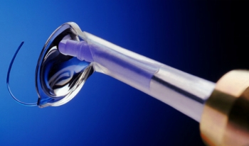
The operation is called phacoemulsification. Using a microsurgical instrument, an incision no larger than 2 mm is made.
Under the influence of ultrasound, the substance turns into an emulsion and is removed from the eye through tubular systems.
The operation is successful in most cases, but complications are possible:
The main disadvantage of the operation is that as a result of removal of the lens, the eye loses its ability to accommodate; it is not capable of focusing images far and near.
If the operation was performed on both eyes, then multifocal glasses are used to achieve focusing of the image on the retinal area.
They have thick lenses and promote distance, near and intermediate vision. Bifocal glasses are also used, but unlike the previous ones, they provide either distance or near vision.
If the cataract was removed in only one eye, then it is advisable to use contact lenses. Since children's eyes are constantly growing, the lenses need to be changed after a while and different sizes selected.
Parents should closely monitor their children's use of contact lenses, as there is a high risk of infection.
After the operation, it is forbidden to rub your eyes for several days, and you cannot swim in pools. Eye drops may be used to moisturize and prevent infection.
Implantation of an intraocular lens
The ideal way to restore vision is surgery to implant an artificial intraocular lens.
The eye begins to function fully, which manifests itself in the form of focusing the image - both far and near.
The operation to implant an intraocular lens is also performed simultaneously and can be combined with cataract removal. The combination is possible in children over 5-6 years old and in adults.
The surgical technique is a sutureless method. An incision of no more than 2 mm is made, and an intraocular lens is inserted using a microsurgical instrument.
The peculiarity of this lens is its small size (otherwise it simply would not fit into the cut). When placed between the pupil and the vitreous body, the lens expands.
Usually, such a lens is implanted in children at least 5 years old.
Since the organ of vision in childhood is in a state of constant development, then Full vision recovery should be expected by adolescence
If the operation is late, amblyopia may develop.. In the preoperative period, due to clouding of the lens, the eye develops incorrectly and “gets used” to not focusing a clear image.
Subsequently after the operation, despite the absence of clouding, the eye also does not focus the image. This phenomenon is called "lazy eye", or amblyopia.
This condition is difficult to combat, so it is advisable to prevent it.
Amblyopia is treated using corrective glasses. The second method is eye activation. To do this, the healthy eye is covered with a bandage, and the patient begins to focus images on the retina.
The longer the patient wears the bandage, the better his vision becomes. There are cases when the sharpness was restored to 100%.
The effectiveness of treatment largely depends on the timing of detection. With early detection and further treatment, vision can be restored. Cataracts are successfully treated in our country.
Prevention of cataracts in children is of great importance. Excessive strain on the eyes should be avoided, fear of injury and compliance with hygiene requirements.
In contact with
Cataract is an eye disease accompanied by clouding of the lens. As a rule, cataracts in children are congenital. According to medical studies, 5,000 out of 100,000 newborns have cataracts; in adulthood, three to four children out of ten thousand.
Cataracts in children can be unilateral or bilateral. It can be treated only by a doctor, based on the severity of the disease and the age of the child.
There are two types of cataracts in children:
Causes
Unfortunately, there are many reasons that cause clouding of the lens of the eye.
Causes of congenital cataracts:
Causes of acquired cataracts in a child:

Symptoms
The manifestation of the disease in a child is influenced by the degree of clouding of the lens, damage to one or both eyes, and the location of the clouding focus.
Even in the maternity hospital, doctors will be able to determine the presence of the disease in a newborn.
At the age of one month, ophthalmologists check the child’s vision during a preventive examination.
Childhood cataracts can be visually recognized if:

Manifestation of cataracts in a child's eye. To enlarge, click on the image.
The first warning sign that a child is developing cataracts may be a change in behavior. For example, a child looks at rattles with just one eye. Schoolchildren begin to study worse, it becomes more difficult for them to focus their eyes and concentrate. Parents who notice such changes should take them seriously and immediately bring their child to be examined by an ophthalmologist.
Note that such symptoms may indicate that the child has other eye diseases. In any case, only a specialist will be able to correctly diagnose the disease and prescribe the necessary treatment.
Diagnostics
As mentioned above, cataracts in newborns can be detected in the maternity hospital. If the child was born healthy, mom and dad should be observant and not neglect visits to doctors.
If the ophthalmologist suspects cataracts, he will instill special drops in the baby that cause the effect of short-term mydriasis (dilation of the pupil). Afterwards, the child's eyes will be examined using special equipment.
If, upon examination, the doctor reveals that the child has no red reflex and clouding of the lens, then most likely he will be diagnosed with cataracts.
Non-surgical treatment
If a child is diagnosed with cataracts, the doctor will prescribe treatment based on the degree of the disease.
In cases where the disease does not prevent the child’s visual system from developing normally, conservative therapy will be prescribed.
For example, a doctor may prescribe drops for the eyes (Quinax, Oftan Katahrom, Taufon). It is prohibited for a child to administer medications at his or her own discretion; prior consultation with an ophthalmologist is required.
Traditional methods
Sometimes, to treat cataracts, they resort to the achievements of alternative medicine:
Surgical treatment
If a cataract becomes a factor interfering with the normal development of the baby’s vision, then it must be removed surgically.
The operation is performed on babies with cataracts no earlier than two to three months of age. Children do not undergo surgery until they are six weeks old because the risk of harm from general anesthesia is too great.
For three-month-old children, the operation is performed using aspiration and irrigation through a very tiny incision.
There are also other types of operations to remove cataracts in newborns:
The operation time is on average two hours. Anesthesia - general. If cataracts affect both eyes, then manipulations are carried out on each eye separately, with a time interval of a week between them.
After surgery, the affected eye is covered with a bandage. The child remains in the hospital for a short time and is then discharged.
Attention: lens implantation surgery can be performed on children over four years of age. Small children are operated on using the irrigation-aspiration method.
Postoperative period
After the patient's eyes have been operated on and the lens has been removed, the doctor will prescribe contact lenses or glasses if both eyes were affected, since the eyes cannot focus on their own.
Also, glasses or lenses are prescribed to children when they were given artificial lenses during surgery. After all, they work like they do - they focus well on distant objects, but close ones will be blurred.
Typically, an ophthalmologist does not prescribe lenses or glasses immediately, but rather some time after surgery. The doctor must explain the rules for caring for lenses and the frequency of changing them.
If your child has had surgery on one eye, the doctor may prescribe wearing a patch on the good eye. You don't need to wear it for long until the operated eye starts working. If previously a diseased eye might not transmit signals from what it saw to the brain, then after wearing such a blindfold the eye will work as expected. The baby needs parental support, the process is not very pleasant.
Please note that after the operation, the child must periodically come to the ophthalmologist for examination.
Summary
Cataracts are a serious eye disease that requires immediate treatment and strict monitoring.
Due to the deterioration of the environment and insufficient control of women over their own health during pregnancy, babies with congenital cataracts are becoming more common.
This disease can also appear after birth. To prevent the development of pathology, you must be attentive to your health and that of your children.
Cataracts in children- symptoms and treatment
What are cataracts in children? We will discuss the causes, diagnosis and treatment methods in the article by Dr. I.V. Elmanov, an ophthalmologist-surgeon with 12 years of experience.
Definition of disease. Causes of the disease
Cataract- clouding of the lens, leading to the development of visual deprivation, that is, deprivation of normal stimulation of the sensory system.
The receptor apparatus of the retina stops working, since due to the cloudy lens, light does not reach the cones, which convert light stimuli into nerve impulses.

The frequency of occurrence of infestation is 1:10,000 newborns. Bilateral cataracts in children are more common than unilateral ones.
All childhood cataracts can be divided into those associated with hereditary anomalies and non-hereditary ones.
Hereditary cataracts:
Non-hereditary cataracts(acquired in utero or in early childhood):
If you notice similar symptoms, consult your doctor. Do not self-medicate - it is dangerous for your health!
Symptoms of cataracts in children
One of the early symptoms of congenital cataracts is the absence of a red reflex during direct ophthalmoscopy.

Congenital cataracts in rare cases are isolated lesions of the lens. More often it is combined with other eye pathologies: amblyopia, nystagmus, microphthalmos, microcornea and other anomalies of the cornea, as well as the vitreous body, choroid, retina and optic nerve.
Changes in the retina and optic nerve of varying nature and severity are the main causes of low visual acuity after extraction of congenital cataracts (removal of the cloudy lens). Most often, combined damage to the retina and optic nerve occurs. Disorders of the optic nerve suggest its partial atrophy and developmental abnormalities (reduction in size, change in the shape of the disc, etc.). On the part of the retina, macular hypoplasia, myelin fibers, central and peripheral dystrophy, and “old” chorioretinal lesions are detected.
![]()
Pathogenesis of cataracts in children
In order to understand the morphology of childhood cataracts, it is necessary to understand how the lens develops during embryogenesis (development of the embryo).

The rudiments of the eye appear in the embryo on the 22nd day of intrauterine development. During the process of organogenesis (4-8 weeks of development), an optic vesicle appears, which invaginates, turning into an optic cup. The interaction of the optic cup and ectoderm stimulates the development of the lens placode. By the fifth week of intrauterine development, the lens vesicle is formed, separating from the surface of the ectoderm. At the eighth week of development, organelles disappear in the primary lens fibers. The equatorial epithelial cells begin to divide, and new cells, being pushed back, elongate and become secondary lens fibers. Where the ends of the secondary lens fibers come into contact in the anterior and posterior poles of the lens, linear “sutures” are formed. The front seams are U-shaped and the back seams are ʎ-shaped.

The final differentiation of fibrillar cells is accompanied by the synthesis and dense packaging of crystallin proteins, which create a transparent, light-refracting environment. The transparency of the lens is also ensured by the programmed disappearance of organelles. At birth, the lens has a spherical shape. Its diameter is 6 mm and as the child grows it increases to 9-10 mm.

An important structural formation associated with the development of congenital cataracts is the pupillary membrane. It is formed as a temporary capillary network. This structure becomes most pronounced at 12-13 weeks of gestation. It nourishes the anterior surface of the lens and then must regress.

The stage of regression of the pupillary membrane is an informative marker of the gestational age of premature infants. At 27-28 weeks, the membrane still completely covers the pupil. At 35-36 weeks of gestation, the pupillary membrane should disappear completely.
Posterior capsular cataracts (retrolental plaque) may also be caused by persistent fetal vitreous vasculature. The hyaloid artery (see picture above) should be completely resorbed (absorbed) by the time the baby is born.
The morphology (structure) of cataracts reflects the age of its onset and determines the prognosis for vision. It can also sometimes be used to judge the etiology of cataracts. The morphology of childhood cataracts is always determined by the anatomy of the lens, its embryology, the timing and nature of the pathological effect that caused it. Acquired cataracts are characterized by a better prognosis for vision than congenital ones.
Some types of congenital cataracts are combined with other eye abnormalities. For example microphthalmos often accompanied by nuclear cataract. Autosomal dominant anterior polar cataracts are often combined with cornea guttata(drip cornea). Wedge-shaped cataracts are characteristic of Stickler syndrome.
Classification and stages of development of cataracts in children
Cataracts in children can be classified into:
According to morphology:

According to the degree of turbidity:
According to the symmetry of the process:
Complications of cataracts in children
If, when cataracts are removed, vision in adults can be completely restored, then a child is initially born with an immature visual system, which is formed before the age of 8–10 years.
Cataracts are a serious obstacle to the normal development of the eye, as they cause sensory deprivation and can lead to the development of persistent obscuration amblyopia, strabismus, nystagmus, and the formation of incorrect fixation.
Diagnosis of cataracts in children
Anamnesis
Includes family history of cataracts in other children (examination of both parents and siblings), corticosteroid use, radiation therapy, and ocular trauma. An examination by related specialists is required to clarify the concomitant pathology. It is important to check with the parents the age when the child was first diagnosed with strabismus, leukocoria, nystagmus, and abnormal visual behavior. It can be informative to analyze photographs taken with a flash when the child’s gaze is directed slightly eccentrically.
Ocular status
Visometry (assessment of visual acuity) can be difficult due to age, although its determination is possible using the optokinetic nystagmus method or using special optotypes (for example, Lea optotypes). Indirectly, visual acuity can be judged by the presence or absence of fixation and tracking of objects.

Indirect ophthalmoscopy allows you to localize lens opacities and examine the fundus.
Ultrasound biometrics necessary for diffuse clouding of the lens, when the deep-lying media of the eye are inaccessible to inspection using the previous method.
Biomicroscopy allows you to accurately assess the morphology of cataracts, but is not feasible in young children. Most often it is performed intraoperatively. It is difficult to sit a child at the slit lamp, but it is necessary to examine his parents. In this case, asymptomatic cataracts may be detected. The study also helps to establish the hereditary nature of the disease.
Tonometry to exclude concomitant pathology - congenital glaucoma.
Assessment of the functional state of the visual analyzer(ERG - electroretinography, VEP - visually evoked potentials of the cerebral cortex) allows you to determine the prognosis for vision after cataract extraction.
Laboratory research
Laboratory tests are less indicated in cases of unilateral cataracts. Such a study is more informative in the presence of bilateral cataracts, as it indicates the systemic nature of the disease.
These days, many parents deliberately refuse vaccination. Therefore, it is necessary to carefully collect anamnesis, and if a child is diagnosed with congenital bilateral cataracts, it is necessary to determine the titer of antibodies to the rubella virus IgM.
Male infants with cataracts, hypotonia, slow weight gain, or slow development should be screened for Lowe's syndrome. To verify the diagnosis, an enzyme analysis (inositol polyphosphate 5‑phosphatase OCRL‑1 in cultured skin fibroblasts) and a test for OCRL‑1 are required.
If you suspect galactosemia should be screened for this disease. Usually it takes place in the maternity hospital, but sometimes it is false negative. More than 20 types of mutations are known to cause autosomal dominant cataracts. Most mutations occur in the crystallin and connexin genes.
In general, we can say that specific tests if a particular congenital syndrome is suspected or if metabolic disorders are suspected are prescribed by a pediatrician or geneticist.
Treatment of cataracts in children
The decision to surgically treat a partially cloudy lens is made only after assessing the morphology of the cataract and the child’s visual behavior. The prognosis for vision correlates more with the density than with the size of the opacities. Nuclear cataracts have a worse visual prognosis. If the large blood vessels of the fundus cannot be distinguished through the central zone of the lens, severe visual deprivation should be expected.
Surgery
Surgical treatment of cataracts that threaten the development of visual deprivation should be carried out in infancy. But most surgeons prefer to wait until the child is 4 weeks old, since there is evidence that cataract surgery before the age of 1 month is associated with a high risk of developing glaucoma. In infants, surgery is performed on both eyes simultaneously, especially in children with underlying medical conditions that increase the risk of general anesthesia. But if surgical treatment is delayed, the interval between operations should be short to avoid the risk of developing unilateral amblyopia. The optimal age for surgical treatment of congenital cataracts is 3-6 months.
Lensectomy and anterior vitrectomy in infants are performed on a closed eyeball. Anterior vitrectomy and posterior capsulorhexis can significantly reduce the risk of repeated interventions. There is no need for phacoemulsification of cataracts in children; phacoaspiration is performed.

The optimal method for correcting aphakia is primary intraocular lens (IOL) implantation. But there are cases when implantation is delayed.
Postoperative management of patients
The main problem of postoperative management of such children is optical correction of postoperative aphakia (lack of lens) and treatment of concomitant amblyopia (“lazy eye”).
Optical stage of treatment
Correction with glasses or contact lenses. Glasses are the simplest and most affordable means of correction. When assigning glasses, you must follow certain rules:

Aphakic glasses are prescribed if a decision has been made to delay implantation. They have optical and cosmetic flaws. For delayed IOL implantation, contact lenses are the optimal correction method.
Pleoptic stage of treatment
A set of therapeutic and training measures aimed at increasing visual acuity. The goal of pleoptics is to create functional equality of the eyes, eliminate amblyopia, and correct incorrect visual fixation.
The main component of pleoptic treatment is occlusion. Occlusion can be:
Auxiliary methods of pleoptic treatment:
Orthoptic treatment
It is aimed at restoring normal retinocortical correspondence, that is, at developing normal binocular vision. This type of treatment is possible only with functional equality of the eyes and central fixation.
After the surgical stage of treatment, the child should be under medical supervision until the age of 12–14 years. During preventive examinations, the refraction and the optical correction required at this stage are clarified, courses of pleoptic treatment are repeated, and complications after the surgical stage are promptly identified and prevented.
Forecast. Prevention
The results of treatment of children with congenital cataracts depend on its early detection, timely (if indicated) surgical treatment and the presence of concomitant pathology.
Continuity in the work of obstetrician-gynecologists of antenatal clinics, neonatologists of maternity hospitals, pediatricians, ophthalmologists of children's clinics and ophthalmologists of specialized medical institutions contributes to the early diagnosis of congenital cataracts and predicting the progression of lens opacities over time.
Timely detection of congenital cataracts in high-risk groups (complicated hereditary history and complicated pregnancy) requires a thorough ophthalmological examination of newborns from the first days of their life - ophthalmoscopy, skiascopy, biomicroscopy in conditions of drug mydriasis (pupil dilation) with an emphasis on the condition of the lens. If a pathology of the lens is detected, the child must be sent to a hospital for a more detailed examination under anesthesia (biomicroscopy, ultrasound and electrophysiological studies) and determination of further treatment tactics.
Severe changes in the lens causing visual deprivation require early surgical intervention. If the lens opacities are minor (partial forms of congenital cataract) and do not require early cataract extraction, then follow-up with an ophthalmologist is recommended:
According to indications, during this period, in order to prevent and treat amblyopia, correction of refractive errors, pleoptic treatment and drug dilation of the pupil are carried out.
Violation of the function of the organs of vision associated with clouding of the lens of the eye is commonly called cataracts. This pathology quite rare, in particular, congenital cataracts in children are diagnosed only in 5 cases out of 100 thousand.
In school-age children and adolescents, the problem occurs much more often, in approximately 4-5 cases out of 10 thousand. Occurs more often in boys.
This is due to the greater susceptibility of the stronger sex all kinds of genetic mutations, which can cause the development of the disease.
The disease requires timely treatment, since as the disease progresses, significant loss of vision is possible, up to its complete loss.
And this means disability, loss of ability to work, significant deterioration in quality of life further.
Concept and characteristics
Cataract is partial or complete clouding of the lens eyes, looks different in appearance, from a small light spot on the lens to a complete change in its shade.
The disease occurs when the chemical balance of substances in the whites of the eyes is disturbed.
Manifests as moderate to significant visual impairment when the child sees the picture as if in a fog. Over time, the pathology progresses, which can lead to complete loss of vision.
What is eye astigmatism in a child? Find out about this from ours.
Causes
Cataracts can be congenital or acquired. Depending on the form of the pathology, the reasons that contribute to its development are different.
|
Congenital |
Acquired |
|
|
|
Classification of the disease
Cataracts in a child may be congenital ( diagnosed immediately after the baby is born, either within a short period of time after birth), or acquired (symptoms of the disease appear several months or years after birth).
Depending on the location, the following types of congenital and acquired cataracts are distinguished:
|
Congenital: |
Purchased: |
|
|
|
There are several stages of development of the disease:
Is it possible to cure myopia in a child? find out right now.
Features of the congenital form

This form of pathology develops in a child even in the prenatal period.
Symptoms of the pathology appear immediately after the birth of the newborn. And these manifestations can be different.
In some cases, there is complete clouding of the lens, leading to significant or complete loss of vision.
Often, against the background of pathology, other eye diseases develop, such as strabismus, nystagmus. Treatment of the disease is carried out surgically.
ICD 10 code – Q 12.0.
Symptoms and signs
Manifestations of pathology depend on the stage of its development. Cataracts in a child can be seen visually, the disease manifests itself as follows:
You will find information on the treatment of farsightedness in children on our website.
Complications and consequences

If cataracts occur, a child needs the help of specialists, competent and timely treatment.
Otherwise, the child may significant or complete loss of vision.
Accordingly, cataracts can cause disability. Other complications are possible, such as:
Non-surgical treatment
Conservative treatment methods have a positive effect if the pathology is successful identify at an early stage of its development when the disease does not cause the child any significant inconvenience and does not interfere with the development of the visual organs.
To treat the disease, special medications are used - eye drops (for example, Quinax, Taufon). The drugs allow you to stop the pathological process and normalize metabolic processes in the eyeball area.
They should be used only as prescribed by a doctor, observing the dosage in accordance with his instructions.
There are also traditional methods getting rid of cataracts at the initial stage of its development:

Surgery
Surgical treatment is prescribed if the pathology is congenital, progresses noticeably, and carries the risk of complete loss of vision. Today, several types of surgical operations are known:
Preventive measures
It is necessary to take measures to prevent the development of cataracts in a child at the stage of planning pregnancy, and throughout its course. Necessary:

To prevent acquired cataracts you must:
Cataract – dangerous disease, which can lead to complete loss of vision and other unpleasant consequences. Often the pathology can be seen with the naked eye, however, to confirm the diagnosis and establish the stage of development of the pathology, it is necessary to consult a specialist.
This is very important, because treatment depends on the form of the pathology, the stage of its development, and the individual characteristics of the child’s body.
Only a doctor can determine these factors and prescribe appropriate treatment after examining a small patient.
Parents It is important to take all measures to promote prevention development of pathology. This must be done before conceiving a child, and throughout pregnancy.
About the causes of congenital cataracts in children in this video:
We kindly ask you not to self-medicate. Make an appointment with a doctor!