Duplex scanning of the vessels of the brain and neck. Transcranial scanning of cerebral vessels
duplex scanning main vessels head and neck (UZDG MAG) allows you to detect significant narrowing or expansion of blood vessels, is used for atherosclerosis, after a head injury, encephalopathy, osteochondrosis of the cervical spine and other pathologies. You can perform duplex scanning of the vessels of the head and neck at the Alternativa Medical Center (Moscow) followed by a consultation with a neurologist.
Duplex scanning of the vessels of the head and neck - what is it?
 Headaches, dizziness, impaired memory and concentration, periodic fainting, decreased hearing or vision - all these and some other symptoms may be a manifestation of impaired blood supply to the brain. Duplex scanning of the main vessels of the head and neck helps to clarify the state of the vascular bed. It combines ultrasound, determination of digital values of vasoconstriction and blood flow velocities, Doppler effect. In the old fashioned way, this study is often referred to as UZDG MAG (ultrasound dopplerography of the main arteries of the head), although modern devices for duplex scanning (this is the device purchased medical center"Alternative" (Moscow)) give a more accurate and detailed information than just an ultrasound.
duplex scanning blood vessels(triplex mode) is the method ultrasound state of the vessels of the head and neck, which allows you to "see" the vessel itself, measure the thickness of its wall, trace the direction of blood flow, and identify its bends. Thanks to the Doppler effect used, color flow mapping is performed.
Duplex scanning of blood vessels of the head and neck - indications for duplex scanning of blood vessels of the head and neck
- head and neck injuries,
- Availability chronic diseases ( , ),
- Takayasu's disease and other vasculitis, determination of different blood pressure on the right and left arms (difference of more than 10 mm Hg),
- Planned heart and vascular surgery,
- Tumors in the head and neck area
- Pathology of vessels of other localization,
- The presence of risk factors for the occurrence of vascular pathology (smoking, sedentary lifestyle).
Duplex scanning of the vessels of the head and neck - the course of the study
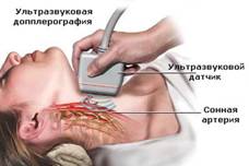
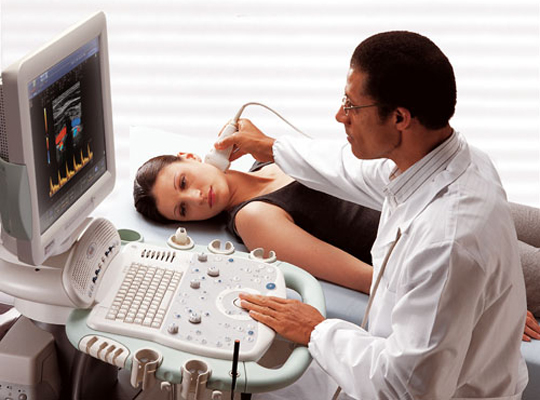
Duplex scanning of the vessels of the head and neck is carried out in the position of the patient "lying on his back". The sensor is moved in the neck of the patient. Special training is not required. For better distribution ultrasound, a special gel is applied to the patient's neck, which, after the examination, is removed with a napkin or towel.
Contraindications: none.
Duplex scanning of the vessels of the head and neck - the advantages of the method
duplex scanning cerebral vessels completely painless and safe for the patient. It is affordable (compared to magnetic resonance imaging) and no less informative.
The possibility of repeated research at certain intervals allows you to evaluate the effectiveness of the therapy.
Duplex scanning of cerebral vessels is a technology for examining cerebral vessels, which consists in combining Dopplerography and standard ultrasound, as a result of which the vessel can be observed in two planes - along and across. Complex use two different ways Ultrasound makes it possible to study the structure of blood vessels and the state of blood circulation in them. Therefore, this technology will be more reliable and informative than ultrasound dopplerography and rheoencephalogram.
The Doppler ultrasound procedure is considered simple for the patient, however, the procedure Ultrasound scanning requires certain skills and equipment. High-quality highly informative equipment for duplex scanning makes it possible to see all the pathologies and changes that occur in the vessels that are hidden under the cranial bones and located in the depths of the tissues. Low-quality equipment not only does not provide complete information about the study, but also misleads the specialist by providing unreliable scan results. Also, vascular scanning requires a high degree of qualification and a course of special training from the specialist who conducts the examination. For the effectiveness of this procedure, the doctor must have knowledge about the features of duplex scanning and be able to correctly interpret the information received during the scan.
Indications for duplex scanning of cerebral vessels
- congenital vascular pathologies and their deformities;
- atherosclerosis and aneurysms;
- traumatic injury and thrombosis;
- angiopathy and inflammatory processes;
- influence pathological processes what happens in the surrounding tissues;
- dyscirculatory anomalies.
Duplex scanning of blood vessels is carried out in the presence of such symptoms:
- the presence of heaviness, noise and pain in the head;
- impaired coordination of movements and unsteadiness of gait;
- dizziness;
- loss of consciousness;
- the presence of numbness and weakness of the limbs;
- speech change.
Patients who have had a stroke and have chronic insufficiency blood circulation in the vessels duplex scanning is performed to determine the state of blood flow and blood vessels. This technology is used to assess and identify secondary vascular deformities in somatic abnormalities. It is also used for the initial determination volumetric formations brain (tumors, hematomas, cysts) and assessment of the state of their blood flow. As a way to objectively monitor the structure of the blood supply, duplex scanning is performed to monitor patients who have undergone vascular surgery or a stroke. Duplex scanning is performed to select conservative method treatment and identification of indications for vascular surgery.
This technology is of great importance for the early detection of vascular anomalies and the prevention of acute damage to cerebral blood flow. The final value in the course of the study will be the determination of the presence of cholesterol deposits in the thickness of the walls of blood vessels. If they are closely soldered to the walls of blood vessels, they do not pose a danger to the patient's health. However, if the plaque can come off vascular wall, then this will lead to clogging of the lumen of the vessels.
Preparation for duplex scanning of cerebral vessels
The implementation of duplex scanning of blood vessels does not require special pre-training. The only requirement for the patient on the day of the upcoming examination will be to refrain from taking substances that affect vascular activity (tea, medical preparations, nicotine and coffee). Failure to comply with these requirements may lead to the receipt of unreliable information during the survey. Medications that cannot be interrupted for a while should be continued. The possibility of a break in the reception should be discussed with the attending physician. Before the actual scanning, the patient needs to remove jewelry and hairpins from the head and neck.
Technique for duplex scanning of the brain
The start of a duplex scan consists in moving the ultrasound transducer over the patient's head to examine the intracerebral vessels or along the neck to study vertebrates and carotid arteries. The sensor sends and captures ultrasound signals from the walls of blood vessels, which the computer processes into an image. The obtained information makes it possible to detect a violation of vascular patency and such causes of its formation as congenital pathologies and vascular loops, narrowing of the lumen and thickening of the vessel walls, as well as the presence of blood clots and vascular junctions.
During duplex scanning, the specialist determines the speed and direction of blood flow, the calculation of indices and the distribution of blood circulation in the vessel, which allows obtaining complete and reliable information about the structure of the vessel under study. To identify the mechanism of vascular regulation, blood flow is studied during exercise tests and functional tests.
Contraindications for duplex scanning of cerebral vessels
Unlike many different diagnostic techniques, duplex scanning has no restrictions and age-related contraindications. Because this procedure may be repeated for as long as it may be necessary to more accurately determine the diagnosis. Conditional contraindications for scanning of cerebral vessels may be unstable and serious condition patient.
With a preventive purpose and at the first stage comprehensive examination vessels of the neck and brain, it is desirable to use non-invasive and safe techniques that allow obtaining preliminary diagnostic information with minimal negative impact on the human body. To do this, triplex scanning is used - a high-tech version of an extended ultrasound examination that helps to assess blood flow in the vessels of the head and neck and identify manifestations of a serious pathology.
What is the triplex method
An echographic study that allows you to diagnose problems in the vessels of the brain consists of 3 types of exposure:
- conventional ultrasound technique;
- transcranial dopplerography;
- color mapping of blood vessels based on the Doppler effect.
Transcranial Doppler scanning is diagnostic study, in which the doctor can see normal or altered blood circulation in the vessels of the brain through the bones of the skull. Modern ultrasound devices equipped with a special Doppler attachment are able to detect vascular pathology in the head, which is actively used in the diagnosis of many diseases in neurological and surgical practice.
The essence of the technique
The standard ultrasound technique helps to assess the anatomy of the organ under study, but is not able to identify vascular disorders and changes in blood flow. Doppler ultrasound should be used to detect problems with the vessels of the head and neck. The method is based on the Doppler effect - this is the reflection of ultrasonic waves from small blood elements moving along the bloodstream.
At duplex scanning the doctor on the monitor screen sees a gray image in the form of graphs, which can be used to suggest the presence of pathology. In a triplex study, using the method of color Doppler mapping, the doctor assesses the situation in the vessels of the brain in the form of a beautiful multi-colored picture, which significantly increases the information content and efficiency of the study. It is the combination of several high-tech methods that allows you to get the best diagnostic result.
What diseases of the cerebral vessels can be detected
Transcranial Doppler sonography is one of the preferred methods for diagnosing vascular pathology of the head and neck. With this method, you can find:
- atherosclerotic lesions of the vessels of the neck and head with the identification of the risk of thrombosis by plaques;
- congenital anatomical changes in the vessels, which can cause dangerous complications;
- inflammatory disorders due to infection with a real risk of thrombosis;
- traumatic damage to the internal structures of the brain with possible trauma to the arteries and veins;
- manifestations of severe neurocirculatory dystonia;
- manifestations of vascular lesions against the background of serious illnesses (diabetes, arterial hypertension).
Often, it is during a Doppler study that a high risk of stroke is first detected, the doctor discovers a tendency to arterial thrombosis or finds some kind of variant.
What diseases of the vessels of the neck can be detected
It is possible to fully assess the state of blood circulation in the head only taking into account the blood flow through the arteries of the neck. The triplex method is actively used to detect pathology in the brachiocephalic vascular bundle, which consists of large vessels- carotid, vertebral, subclavian and brachiocephalic arteries. Doppler ultrasound of the BCA can identify the following life-threatening problems:
- arterial thrombosis by atherosclerotic plaques;
- injury to the vascular bundle in the neck.
A triplex study of BCA should be performed in all cases when cerebral disorders occur, there is a risk of stroke, or typical symptoms of blood flow disorders in the vessels of the head appear.
Who is shown the study
The undoubted advantage of transcranial Doppler scanning is absolute safety for humans. This technique should be applied in the following cases:
- with frequent recurring severe pain in the head;
- with unexpected problems with vision and hearing (tinnitus, sharp drop visual acuity, auditory and visual hallucinations);
- with dizziness and fainting;
- at high blood pressure accompanied by nausea and vomiting.
Preventive examination of the vessels of the head and neck using Doppler ultrasound should be carried out:
- people with chronic long-term diseases (diabetes, obesity, hypertension, heart disease);
- people who have had at least 1 episode of stroke or any variant of the disorder in the past cerebral circulation;
- people with a long history of smoking;
- people who have undergone any type of surgery on the vessels of the neck and head.
Sometimes even a severe migraine should be an indication for a complete and comprehensive examination with a diagnosis of the condition. vascular system brain. In this case, do not worry - ultrasound scanning with dopplerography is absolutely safe method having no contraindications for the study.
In most cases, when symptoms of a blood flow disorder in the head occur, the doctor chooses the examination method that will help to quickly make a diagnosis. At the same time, safety for the patient is always taken into account, therefore, Dopplerography is used at the 1st stage. To clarify the diagnosis and obtain more complete information about the vessels of the head and neck, MRI or CT is used.
Health problems associated with the state of the blood vessels of the neck and head require timely studies of arteries and veins. Any disorders that cause discomfort, leading to pain and disease should be detected on early stage. For the most accurate and detailed study, triplex scanning of the vessels of the neck and brain can be performed. This research aims to overall picture describing the current state of the veins and arteries, the need for their treatment, possible problems in future.
Description of the procedure
Before carrying out any research procedure, each patient wants to know what it is and why it is performed.
Triplex scanning includes general analysis vessels, which allows you to obtain the following information:
- their structure (including integrity, the presence of a narrowing or expansion of the site, any modifications);
- patency of the vessel (the presence of plaques, blood clots, their current position and size, the risk of further movement and occlusion of a vein or artery);
- blood flow (corresponding to norms, problems with its movement from the heart to the head and neck or vice versa).
A detailed analysis allows you to identify any deviations caused by the development of the disease or the prerequisites for its occurrence. This type of scanning includes three operating modes at once: ultrasound, doppler, color mapping. The latter mode allows you to quickly visually assess the blood flow in the veins and arteries. It consists in showing black and white pictures(section of the studied segment) and overlays of colored zones (blood flow) on it.
The color of the blood is carried out according to the registered flow velocity. Such an examination of the vessels of the neck and head greatly simplifies the possibility of getting acquainted with the state of the region and the correct operation of the vessels.
Indications for carrying out
Damage to arteries and veins as a result of trauma, their destruction due to disease, or occlusion can lead to the appearance of different symptoms or multiple signs of disease. Therefore, in case of any suspicions (heredity, features of the structure of blood vessels), extended examinations should be started immediately. They will help warn dangerous diseases and exclude their development.
A triplex scan of the vessels of the head and neck is mandatory when the following symptoms appear:
- constant drowsiness, fatigue, impotence;
- dizziness (up to fainting);
- visual enlargement of the veins;
- feeling unwell (in rare cases, it may be accompanied by hallucinations);
- periodic numbness of the fingers or limbs (as well as their redness);
- problems with memory or speech, perception of information;
- the appearance of "blinking" dots before the eyes, vision problems or its complete disappearance (in one of the eyes);
- the appearance of cramps in the limbs;
- insomnia, tinnitus;
- pulling or sharp periodic pain in the neck, head, frequent migraines.
An urgent appeal to specialists must be performed in the presence of any diseases of the vessels of the neck or head. Such scheduled examinations allow you to closely monitor the change in the state of the structure of the vessels, possible complications. Examinations are mandatory if the patient has: atherosclerosis, osteochondrosis in the cervical region, cerebrovascular accident, predisposition to stroke.
The presence of aneurysms in the neck or head may also be an indication for a triplex. This will help to avoid cases when, as a result of an advanced disease, problems with the veins and arteries caused the death of the patient.
Advantages
Indications for scanning indicate the occurrence anxiety symptoms, which may be a consequence of the disease or its development. A timely study, properly selected treatment can prevent a vascular crisis and other diseases that are dangerous to the health and life of the patient.
The main advantages of triplex are:
- Detail of received researches.
- Efficiency of conducting (simplicity of studying the data and a detailed result).
- Fast (on average, scanning can take from 30 to 50 minutes).
- Safety (ultrasound exposure is not negative and does not affect the patient's condition or health in any way).
- Universality (suitable for people of all ages, regardless of whether they have diseases of other body systems).
Triplex examination of the vessels of the head and neck differs from other types of scanning and obtaining all possible types of data that will help in as soon as possible determine the optimal treatment.

Features of patient preparation
The simplicity of the study in modern centers and its safety eliminate the need for special patient preparation. The only thing that is not recommended to do before the scan is to take drugs that can affect vascular tone.
- avoid smoking before the procedure;
- refuse to take tonic and alcoholic drinks;
- Do not drink coffee or other coffee drinks.
In addition, patients should be psychologically prepared for the study. In their work, specialists use only a special apparatus that allows them to inspect vessels, a gel. Awareness complete security research will allow the patient to relax, and the doctor himself to conduct a detailed analysis of the condition of the veins and arteries. The doctor will examine cervical region and will assess the state of the vessels of the head. Further, after literally 20-40 minutes, an accurate and detailed result of the study will be obtained.