Duplex scanning of the great vessels of the head. Duplex of head and neck vessels: how the procedure works, pros and cons
Today, triplex scanning of blood vessels (angioscanning) is an accessible and reliable method of diagnosis using ultrasound machine. As a result of the study, an image of the arteries and veins is obtained, and the characteristics of the blood flow in them are also traced. This diagnostic Head and neck therapy is based on the process of transmitting ultrasonic waves into tissue through the bones of the skull and receiving an echo signal. The doctor moves a special sensor in the area where the vessels necessary for examination are located and an image is displayed on the monitor in real time.
What is the purpose of triplex scanning?
Angioscanning of the head and neck allows us to identify the following abnormalities in functioning: circulatory system: blood clots, pathological tortuosity, aneurysms, cerebral blood supply disorders, atherosclerotic plaques. In the latter option, it is even possible to assess the degree of vasoconstriction, which is especially important if the patient is about to undergo surgery.
Timely angioscanning of the vessels of the head and neck will help avoid the development of many diseases of the circulatory system. The reliability of the research results will depend on the qualifications and experience of the doctor conducting it.
Angioscanning allows you to obtain accurate information about the state of blood flow, vessels of the head and neck, the risk of developing various pathologies, many of which can lead to disability or fatal outcome. Thanks to the data obtained, it will be possible to choose more effective method treatment and most accurately predict its outcome.
Significant advantages of angioscanning
- There are no contraindications to the study.
- Does not involve radiation exposure, since it is not related to ionizing radiation.
- Good diagnostic value.
- Can be done many times without any harm to health.
- Does not cause discomfort or any pain.
- No special preparations are required, including discontinuation of medications.
Duplex or triplex study

Today, many patients are confused by various formulations medical research, which are prescribed to them, and do not know what is best to do and when. For example, triplex or? This can be misleading for many, although, in fact, there are no fundamental differences between duplex and triplex scanning of the brachiocephalic arteries. In the first case, the condition of the arteries is assessed simultaneously in two ultrasound modes, in the second - in three. Modern ultrasound systems are equipped with all necessary operating modes. They are chosen by the doctor based on physiological characteristics and suspected pathologies in each individual case.
Advice: Properly performed triplex scanning will eliminate any need for expensive studies in which a contrast agent is introduced, which can cause the appearance of allergic reactions.
Triplex examination of the vessels of the head and neck was created on the basis of duplex scanning, and therefore is more informative. It is supplemented with color Doppler mapping. That is, we can distinguish three main functions of such a study of the vessels of the head and neck:
- Blood flow assessment.
- Study of vascular anatomy.
- Assessment of the patency of arteries and veins in color mode.
In what cases is triplex scanning performed?
Angioscanning of the head and neck is usually prescribed for complaints of migraines, headaches, dizziness, and dizziness. blood pressure, injuries, after surgical interventions. In addition, if necessary, the lower and upper limbs, a triplex study is also being conducted.
As you can see, this type of ultrasound tests is widely applicable to identify various violations the entire bloodstream. In terms of price and reliability of the results obtained, it is a more optimal option, therefore it is especially popular among angiosurgeons and their patients.
Attention! The information on the site is presented by specialists, but is for informational purposes only and cannot be used for self-treatment. Be sure to consult your doctor!
Duplex scanning vessels of the head allows a specialist to analyze the condition blood arteries, their geometry. The procedure makes it possible to examine existing deformations, detect transcranial deviation, branching of arteries, and their length.
After passing the examination, it is determined:
- Elasticity
- Rigidity
- Integrity
- Wall thickness
- Structure violations
- Intraluminal formations
- Echogenicity
- Length
- Change in arterial diameter
- Luminal patency
Duplex scanning of cerebral vessels makes it possible to establish an accurate diagnosis when the patient is diagnosed with:
- Inflammation of capillaries
- Arterial injury
- Diseases of the walls of blood vessels
- Types of angiopathy
- Vascular dystonia
- Discirculatory encephalopathy
Duplex scanning is prescribed for diseases:
- Endarteritis
- Diabetes
- Aneurysm
- Varicose veins
- Vasculitis
- Vascular trauma
- Phlebothrombosis
- Thrombophlebitis
What arteries are examined?
Duplex scanning of cerebral vessels is done to study the patency of blood vessels. The ultrasound examination provides very accurate results about the condition of human blood arteries.
An ultrasound helps to study the condition of the main arteries and check the speed of blood flow. The display clearly shows the vessel surrounded by tissue. Such diagnostics allows us to determine the causes of poor arterial patency. Visually you can see:
- Blood clots
- Plaques
- Thickenings
- Ornateness of blood vessels
Very good results gives a triplex study of the body. The display shows a colored vessel, and its color depends on the speed of blood flow.
Preparatory operations for the examination
Duplex scanning of the vessels of the head and neck, as well as their ultrasound examination, is done without special preparation. Before starting the study you should not consume:
- Energetic drinks
- Alcohol
- Tobacco
Certain types of medications can also distort ultrasound results:
- Betaserk
- Vinpocetine
- Cynarizine
- Fezam
- Antihypertensive drugs.
Before the examination begins, there should be no jewelry on the neck or head. After completing such an examination, you must wash your hair.
Process technology
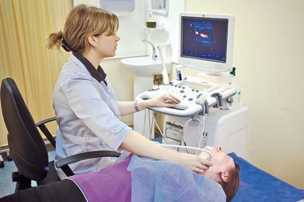
Carrying out Doppler examination, as well as ultrasound, is done according to the same principle. The patient should lie on his back. A hard pillow is placed under the head; it can be replaced by a bolster.
 The patient turns his head, allowing the neck to be examined. A special gel is applied to the skin to facilitate the movement of the transducer. Thus, the doctor checks the condition of the arteries and takes the necessary measurements.
Diagnosis of extracranial cerebral vessels is carried out through the cranial bones. The sensor is located on the head, in the area of the temporal areas. These areas are covered with a water-soluble gel, which helps to obtain the most objective ultrasound results.
In addition to a visual examination of the arteries, the doctor makes special functional tests, for example, asks the patient to hold his breath. In this way, he can check whether there are disturbances in autonomic regulation.
Need for scanning
A study of the condition of cerebral vessels using the duplication method is prescribed when signs of chronic cerebrovascular insufficiency appear. This also includes the appearance of frequent headaches.
The doctor prescribes such an examination before performing operations related to the presence of cardiac arterial pathology. First of all, this concerns IHD.
Transcranial scanning of the arteries is performed before the stenting procedure. Ultrasound examination is prescribed if there is a risk of cerebrovascular pathologies. The reason may be  become:
- Smoking
- Arterial hypertension
- Obesity
- Hyperlipedemia
- Diabetes
Results of extracranial work studies great vessels help the doctor determine the type of blood flow, the existing speed, and identify defects in the filling of the arteries.
Ultrasound of the venous bed makes it possible to determine:
- Anatomical structure
- Patency
- Diameter
- Ornateness
- Blood speed
- Intraluminal formations
After checking the vessels, the ultrasound report does not show any digital data. Doppler scanning of arterial vessels allows for digital analysis, and it becomes possible to compare study data with norms.
Signs of vascular pathology
Ultrasound helps determine pathological changes venous system, give her accurate diagnosis. Most often detected:
- Arteriovenous malformations
- Aneurysms
- Varicose veins
If non-stenotic atherosclerosis occurs, disturbances in the functioning of the main arteries, a decrease in the lumen, and thickening of the arterial walls are detected.
Identification of plaques indicates stenosing atherosclerosis.
Ultrasound can detect an inflammatory process, changes in the thickness of the walls of blood vessels, and the appearance of vosculitis.
Temporal arthritis is characterized by diffuse thickening of the arteries located in the temporal region, low echogenicity is determined. In case of prolonged inflammatory process, the appearance of atherosclerotic lesions is possible.
Diabetes mellitus is characterized by the appearance of signs of macroangiopathies.
Anomalous phenomena are very common in the area vertebral arteries, so-called hypoplasia. In other words, the diameter of the artery decreases. Sometimes it becomes less than 2 millimeters.
Ultrasound symptoms of hypoplasia depend on its severity. Ultrasound records the appearance of abnormal phenomena at the junction of the vertebral main artery with the vessels of the cervical vertebrae. Typically, such an anomaly does not affect hemodynamics.
Ultrasound shows existing extravasal compression, that is, compression of the vessel walls. The reason for this compression of the neck vessels is:
- Inflammation of the thyroid gland
- Severely enlarged lymph nodes
- Tumors
- Osteophytes
Benefits of Duplex Scanning
When such a procedure is carried out, it is determined:
- The state of blood flow in the vessels of the brain
- Are revealed early stages changes in the structure of blood vessels, such as atherosclerosis
- Vascular patency
 The main advantage of duplex scanning is the ability to diagnose early signs of the disease, when characteristic clinical symptoms have not yet appeared.
Such a scan helps to identify existing pathologies associated with the functional functioning of the blood flow. Examination ultrasound diagnostics dispenses with the use of an X-ray machine, there is no radiation at all, such diagnostics can be carried out many times.
Ultrasound is a non-invasive method. In other words, no procedural examination is required, no application is required. special medicines. The occurrence of allergic reactions is completely eliminated, and there are no side effects of any kind.
Today, duplex scanning of the vessels of the brain and neck is considered the most effective way a study that allows the doctor to obtain maximum information about the condition of the human blood vessels.
For a long time, this type of examination of the human body, its blood vessels in the brain, has been considered the most objective and completely safe. Of course, the result of the study largely depends on the qualifications of the specialist deciphering the study. Errors during such verification are not allowed.
Thanks to duplex scanning, many patients avoided the occurrence of dangerous diseases, were able to improve the condition of their blood vessels in time.
After a duplex scan, the doctor can prescribe the most appropriate treatment, after which the person can live a normal, full life.
Qualitative diagnosis of diseases of the arteries and veins in the brain and cervical spine spine makes it possible to detect dangerous pathologies, life threatening patient, long before their manifestation clinical signs and begin timely and adequate treatment. Duplex scanning of the vessels of the head and neck is a non-invasive modern technique, which is a type of ultrasound, with which you can visualize each artery and assess the speed of blood flow in them. In this case, the tissues that surround the vessels are visible.
Ultrasound duplex scanning (USDS) is based on the practical use of the properties of an ultrasonic wave, which, when reflected from red blood cells moving in vessels, makes it possible to obtain an image of the vessel being examined.
This technique allows you to effectively examine both extracranial (cervical) and intracranial (located inside the brain) veins and arteries.
The doctor talks about the diagnosis ultrasound diagnostics Irina Gennadievna Maslova:
Duplex examination is based on two diagnostic technologies. Firstly, it is ultrasonic radiation configured in pulse mode, due to which an image is displayed on the monitor. Studying it, the specialist evaluates the structure of the tissues and examines the vessels. Secondly, the Doppler effect, which involves assessing a moving object, which is the blood flow, by reflected ultrasound on different areas veins.
When an image is fed to the monitor, a two-dimensional image is formed along and across the arteries.
The use of such a non-invasive diagnostic method is necessary to obtain information reflecting changes in the condition of blood vessels, as well as their effect on blood flow. Transcranial duplex vascular scanning (TCDS) allows for valuable research to detect changes that occur in the tissues and arteries of the brain, making it possible to diagnose brain lesions and establish control over the flow pathological process.
Using this method, the following information is obtained:
- Condition of the walls of blood vessels in the neck and head;
- Condition of vascular tissue;
- Presence of stenosis and assessment of its severity;
- Disturbances in the flow of the bloodstream;

- Anatomical features circulatory system in the head and neck area;
- The presence of formations inside the vascular canals;
- Deviations associated with changes in the course of the arteries.
To obtain the most reliable information, experts use modern technology, with which you can conduct a so-called color scan (CDS) - the ability to display a color sketch of the blood flow on the monitor, which facilitates the process of determining its speed in various areas.
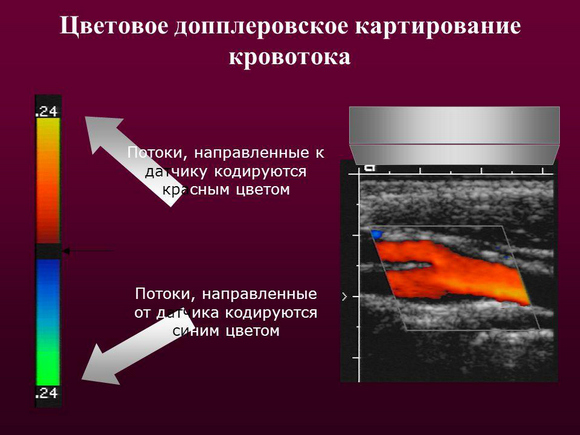
Color Doppler mapping (CDC) is a subtype of manipulation based on the Doppler effect. In this case, data on the speed of movement of structures is displayed in different colors. In particular, the color red determines the speed of blood flow towards the sensor, and the lighter the shade, the slower the speed. The blue color in the study indicates the speed of blood flow in the direction from the sensor. Using this technique, not only a specific vascular pathology is determined, but it is also possible to distinguish a benign process from a malignant one, and to identify the tumor’s tendency to further growth and spread.
Indications for use. What pathologies does the procedure reveal?
Intracranial duplex, which involves the study of veins and arteries located in the cranial cavity, is indicated for patients who have the following complaints:
- Regular headaches;
- Dizziness;
- Noise in the head or ears;
- Fainting;
Ultrasound diagnostics doctor Murat Medzhidovich Nagaplev talks about the indications for the examination:
- Manifestations of inappropriate behavior;
- Weakness and numbness of the limbs;
- Violation visual functions;
- Poor coordination combined with unsteadiness, with an uncertain gait;
- Deviations in speech production or understanding.
TKDS reveals circulatory disorders in the head area. It is prescribed when the following pathologies are detected:

- Intracranial hypertension;
- Lesions of intracranial vascular canals.
Scanning of extracranial vessels located outside the skull and supplying blood to the brain is carried out when the following pathological signs appear in the patient:
- Impaired ability to remember information;
- Inability to concentrate on anything;
- Dizziness, intense headaches;
- Coordination problems.
The arteries and veins of the cervical spine should also be examined if it is necessary to perform an operation involving intervention in the vessels of the heart or directly in the muscle structure, as well as when identifying pathologies of the neck organs that can lead to compression of the vessels located in this area.
Duplex scanning of the vessels of the brain and cervical spine is a procedure that should be performed routinely (once a year), even in the absence of any complaints, in the following cases:
- If the patient is over 40 years old (men) or 45 years old (women);
- If you have close relatives who have diseases such as diabetes, hypertension, ischemic disease;
- If the patient is an experienced smoker;

- With previously conducted surgical interventions on the spinal cord or brain;
- In case of stroke or disorder cerebral circulation in the anamnesis;
- In the presence of .
Ultrasound duplex scanning is used to diagnose the following pathologies:
- Venous thrombosis, thrombophlebitis, thromboembolism pulmonary artery;
- Anomalies and injuries of veins;
- Varicose veins;
- Aneurysm;
Rehabilitation doctor Sergei Nikolaevich Agapkin tells more about cerebral aneurysm:
- The degree of blood flow deficiency;
- Atherosclerosis;
- Ischemia;
- Angiopathy;
- Vasculitis.
A method such as ultrasound is characterized by high information content, painlessness, and the absence of harmful effects on the body, since the manipulation does not require equipment with radiation.
How is diagnosis carried out?
Diagnosis of the main arteries of the head is a safe measure, which, if necessary, can be prescribed to pregnant women or a child. Relative contraindication to the procedure is general serious condition patient or the presence of diseases that make it impossible for the patient to accept horizontal position, in which the procedure is carried out.
Examination of the large cervical arteries
The examination lasts no more than half an hour. Any preliminary preparation no scanning required. Only one thing is required from the subject - not to take substances or drugs the day before that affect vascular tone and distort big picture their condition. These include caffeine, nicotine-containing substances, energy and alcoholic drinks. You should not visit the sauna or bathhouse before duplex scanning.
Duplex scanning is carried out as follows:
- The patient is in a supine position. The specialist fixes the head so that it is in an elevated position, for which a cushion is placed under the neck. The head is turned in the direction opposite to the area in which the study is being carried out;
- The specialist uses a sensor to trace the area where the vessels are located. Previously, to facilitate the examination, a gel with a special composition is applied to the surface of the skin. The resulting image is fed to the monitor;

- The examination begins with diagnosis carotid artery to the entrance to the skull, examining it in different planes;
- When examining the brain, areas such as the occipital bone, temporal and supraorbital regions, and the junction of the occipital bone with the spine are examined.
During the duplex of the vessels of the head and neck, functional tests can be performed to study autonomic regulation. For these purposes, the specialist may ask the subject to hold his breath, cough, and slightly change his body position.
As for the cost of the procedure, it is not too high and, depending on the level medical institution and the city in which it is located ranges from 2300 to 4000 rubles.
Results and their interpretation
The condition of the arteries and veins of the head and cervical region is assessed using such indicators as the thickness of the vessel wall, the nature and speed of blood flow, the ratio between the minimum and maximum speeds.
The level of Doppler indicators of the condition of arterial vessels is expressed in numbers. Thus, the norm for artery wall thickness is from 0.9 to 1.1. Normal indicator maximum systolic velocity should not exceed 0.9, peak velocity in diastole - less than 0.5.
Decoding the results allows you to obtain the following signs of vascular pathology:
- An increase in wall thickness and a narrowing of the artery by less than 20% indicates atherosclerosis;
- Diffuse change wall thickness indicates vasculitis;
- The presence of a fistula between veins and arteries is a sign of malformation.
A lecture on the symptoms and treatment of vascular atherosclerosis is given by general practitioner Yuzef Viktorovich Krinitsky:
The examination protocol allows the specialist to determine the most early signs disease, even before manifestation clinical symptoms.
The advantages of duplex scanning of the vessels of the head and neck are that it is a reliable method that does not require the introduction of any substances into the patient’s blood, as well as exposure to x-rays which may adversely affect your health. Diagnostic method has no contraindications. In addition, the price of the procedure is not too high, which will allow any patient to quickly and accurately determine the condition of the vessels of the neck and head.
Duplex scanning of the vessels of the head and neck is one of the most modern methods of vascular research. From this article you will learn about the essence of the procedure, indications for its purpose and information content.
Vascular duplex scanning or vascular duplex is a special type of ultrasound or sonographic examination designed specifically to study vascular structures and the nature of blood flow in them. This method is different from the usual ultrasound examinations a combination of two main methods:
- The usual B-mode ultrasound is the same gray-white picture depicting organs and tissues in a planar mode. In duplex mode, depending on the tilt and rotation of the sensor, vessels look like longitudinal or transverse sections. In this mode, you can examine their course, measure the diameter, evaluate the lumen, the presence of blood clots or other inclusions in them. Cutting-edge sensors can even help you see the image layer by layer vascular wall- like a section in a microscope.
- Doppler mode is a type of ultrasound examination based on recording the flow of moving blood particles in the vessels. Using the Doppler effect, you can record the fact of blood flow, evaluate its intensity, direction of blood movement, measure its speed, resistance indices and other important indicators.
Procedure for duplex scanning of neck vessels
Duplex examination of all vessels takes place using similar principles. human body. The greatest importance in modern medicine has duplex scanning of arteries and veins lower limbs, hearts and his large vessels, as well as the vessels of the head and neck. It is this latter study that we will talk about in a little more detail.
Why do head and neck vessels duplex? The fact is that the brain, its structures and vascular system have always been a stumbling block in diagnosis. The brain is securely covered by the cranium, so just a few decades ago the only objective method his research was X-ray studies. In addition to X-rays, various neurological tests were used to evaluate brain function and indirect signs cerebrovascular accidents. Modern techniques successfully study the vessels of the head and neck that supply the brain that are accessible to research. Thus, by assessing the blood flow in these feeding trunks, one can indirectly judge the similar blood flow in the brain tissues.
The brain is supplied with blood from the branches of the carotid artery, vertebral arteries, subclavian arteries and brachiocephalic trunk, which mutually replace each other and form the circle of Willis. The more traditional and generally accepted name for all these arteries is the brachiocephalic arteries, or BCA. Accordingly, duplex scanning of the vessels of the head and vessels of the neck is more often called. It is prescribed by neurologists or therapists, and performed by ultrasound doctors.
 Circle of Willis
 Indications for the study
Let us define a list of conditions and diseases that can and should be studied by this method:
- or suspicion of it. Such suspicions arise with an unfavorable cholesterol and triglyceride profile, excess body weight, hereditary history, and neurological signs of cerebral blood flow suffering.
- Endoarteritis of the vessels of the neck and brain - most often autoimmune disease, affecting the wall of the arteries of the human body.
- Aneurysms and other vascular malformations in the cranial cavity - already diagnosed based on the results of previous ultrasound, angiography, computed tomography or suspected based on clinical symptoms or patient complaints.
- Vasculitis is a wide group of inflammatory vascular diseases, including autoimmune.
- Condition after surgical treatment vessels of the head, neck or brain.
- Conditions accompanied by external vessel syndrome. Such conditions include compression of vascular trunks by pathological foci, hematomas, bone fragments and tumors.
- Vascular thrombosis or suspicion of them.
- Head and neck injury.
- Ultrasound of the BCA can be prescribed in doubtful cases and when it is unclear clinical picture: memory impairment, pathological drowsiness, convulsions, mental changes, unclear headaches, dizziness, loss of consciousness, deterioration of vision and hearing.
 The main symptoms of vasculitis - an autoimmune vascular disease
 Pros and cons of the method
Like any diagnostic method, duplex scanning has positive and negative sides. Let's start with the pros:
- Complete security. Medical ultrasound does not have any damaging or disfiguring effects on the human body, therefore this study Can be performed on young children and pregnant women.
- Painless and non-invasive. The duplex scanning procedure is completely painless and does not bring any discomfort to the patient.
- High diagnostic accuracy. Considering that the doctor sees the vessel being examined in two projections, while simultaneously recording the blood flow in it, the information content of such studies is comparable to angiography.
- Relative ease of research. Ultrasound scanning takes 20 minutes at most, does not require bulky equipment or the help of additional medical personnel, but requires the deepest knowledge of the anatomy and physiology of blood vessels from the research physician.
- There are no contraindications or complications of the procedure - the study can be prescribed to absolutely all categories of patients, and throughout history no serious consequences have been recorded.
- No special preparation for the study is required, such as diet, cleansing enemas, taking certain medications and administering solutions.
Let us list the disadvantages of the technique:
- The relative cost of the method. The fact is that duplex studies require special ultrasound machines, which are often unavailable to small clinics and government agencies.
- Possibility of research “here and now”. This general disadvantage of all ultrasound examinations, since the “picture” can only be assessed in real time, in motion, and not from frozen images.
- Narrow study area. It is important to understand that scanning the brachiocephalic vessels only indirectly indicates blood circulation in the brain. Through cranium Ultrasound waves cannot penetrate an adult, so the brain itself and its vascular system are hidden from the researcher.
How the research is carried out
The procedure for duplex scanning of the vessels of the head and neck is quite simple and takes no more than 30 minutes:
- The patient arrives at the appointed time and sits on the couch. The examination can be carried out with the patient lying on his back or side, sitting or semi-sitting. No special preparation is required for the procedure.
- The doctor applies a special gel to the sensor and the patient’s neck.
- Next, the doctor easily moves the sensor along the side of the neck. Sometimes a diagnostic specialist may ask the patient to change his body position, cough, strain, or hold his breath.
Today, ultrasound research methods that can be used to diagnose vascular pathology of the brain are presented:

Duplex scanning of the vessels of the head and neck is recognized as the gold standard for diagnosing angiological pathology in this area. What is the essence of this method? How does it differ from ultrasound and ultrasound? Who is indicated for this study and how to prepare for the procedure? We will answer these and many other questions in the article.
Review from our reader Victoria Mirnova
I’m not used to trusting any information, but I decided to check and ordered a package. I noticed changes within a week: constant pain in my heart, the heaviness, pressure surges that tormented me before receded, and after 2 weeks disappeared completely. Try it too, and if anyone is interested, below is the link to the article.
What is a duplex?
Duplex scanning combines the advantages of classic ultrasound and Doppler.
 This research makes it possible to:
- obtain an image of the vessel, evaluate its morphology (something that can be done with regular ultrasound);
- visualize the blood flow in the lumen of the vessel, evaluate its direction, intensity, as well as a number of other characteristics (what ultrasound scanning allows).
Thus, it is possible to evaluate two basic parameters– structure and function. In addition, duplex allows visualization of atherosclerotic plaques, thrombi and emboli of various origins, pathological tortuosity blood vessels, thickening or thinning of vascular walls.
Modern ultrasound machines, which provide duplex scanning, produce color images.
The resulting picture is displayed in the form of a cartogram, which contains information about the speed, direction, and intensity of blood flow. This option The technique is called color Doppler scanning (CDS).
 The extensive possibilities revealed by this method in the diagnosis of angiological pathology determine wide range indications for the study.
Using this method of ultrasound diagnostics, you can examine almost any vessels human body. Since this article is about the head and neck duplex, below we will provide a list of vessels in this area that are most often examined using this technique.

These are the main groups of vessels that are available for duplex scanning of the corresponding area.
The method of transcranial Doppler scanning (TCDS) does not lose its relevance. This study allows you to evaluate blood flow in the same vessels that are accessible to the duplex. The disadvantage of the method is the inability to assess the morphology of the vascular bed. Therefore, transcranial Doppler is used only when duplex is not possible.
To clean VESSELS, prevent blood clots and get rid of CHOLESTEROL, our readers use a new natural drug recommended by Elena Malysheva. The product contains blueberry juice, clover flowers, native garlic concentrate, rock oil, and wild garlic juice.
Indications for the study
Let's start with the cases in which routine duplex scanning of the head and neck area is necessary. Duplex scanning of cerebral vessels is indicated for:

Separately, cases should be analyzed when the patient is bothered by certain symptoms, the diagnosis has not been established and an ultrasound examination is recommended. Symptoms that indicate the need for a duplex of the head and neck vessels include:

In the above cases, duplex scanning will reveal pathology and establish a diagnosis. The efficiency of the study increases significantly when using the CDS mode, which was already mentioned above.
Several nosological units can be identified, in the diagnosis of which the method of duplex scanning of cerebral vessels plays a decisive role. This:

Thus, modern techniques Ultrasound allows you to diagnose many different pathological processes. And an accurate diagnosis is the key the right choice patient management tactics.
How is the research conducted?
Carrying out this type of ultrasound (including with CD) does not require special preparation of the patient. The only thing the attending physician can ask is a refusal to use substances that may distort the real picture. We are talking about substances that significantly affect vascular tone.
Many of our readers actively use the well-known method based on Amaranth seeds and juice, discovered by Elena Malysheva, to CLEAN VESSELS and reduce the level of CHOLESTEROL in the body. We recommend that you familiarize yourself with this technique.
 Therefore, on the eve of the duplex, the head and neck area should be avoided:
- nicotine-containing substances;
- caffeine-containing substances;
- alcohol and energy drinks.
If the patient regularly takes medications that affect vascular tone, then before performing this type of ultrasound, he should discuss the issue of medication regimen with his doctor.
All types of ultrasound of the vessels of the head (including duplex with CDS) are carried out according to a single scheme. The patient is in a lying position, with a special cushion (or pillow) under his head.
The neck should be free and the head should be turned in the direction opposite to the side of the study. A special gel is applied to the skin, along which the sensor moves. The doctor places the sensor at certain points, which makes it possible to visualize the required part of the vascular bed.
Duplex is simple, non-invasive and absolutely painless method. At the same time, this study is extremely informative and effective.
About the information content of this method
In some cases, the duplex of cerebral vessels makes it possible to solve diagnostically complex tasks. Below we give several examples in which cases we can resolve the issues of diagnosis and choice. therapeutic tactics is not possible without using duplex scanning:

From all of the above, it becomes clear that today duplex scanning of cerebral vessels plays an important role in diagnosing diseases of neurological and angiosurgical patients. Duplex provides high quality diagnosis, and therefore subsequent treatment.
Do you still think that it is completely impossible to RESTORE blood vessels and the BODY!?
Have you ever tried to restore the functioning of your heart, brain or other organs after suffering pathologies and injuries? Judging by the fact that you are reading this article, you know firsthand what it is:
- often occur discomfort in the head area (pain, dizziness)?
- You may suddenly feel weak and tired...
- is constantly felt high blood pressure…
- there is nothing to say about shortness of breath after the slightest physical exertion...
Did you know that all these symptoms indicate INCREASED CHOLESTEROL levels in your body? And all that is necessary is to bring cholesterol back to normal. Now answer the question: are you satisfied with this? Can ALL THESE SYMPTOMS be tolerated? How much time have you already wasted on ineffective treatment? After all, sooner or later the SITUATION WILL GET WORSE.
That's right - it's time to start putting an end to this problem! Do you agree? That is why we decided to publish an exclusive interview with the head of the Institute of Cardiology of the Ministry of Health of Russia, Renat Suleymanovich Akchurin, in which he revealed the secret of TREATING high cholesterol.