How is an ultrasound scan of the veins of the lower extremities performed? How effective is ultrasound of the vessels of the lower extremities? Preparation and how to do it
Before signing up for the procedure, people wonder: how to do ultrasound of veins lower limbs ? Many people worry about pain or the long preparation phase. No one wants to be turned away from a clinic because the preparation has not been completed. But here the principles are different and dieting and no meals are not required, as is the case with checking the gallbladder or liver.
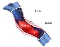
An ultrasound examination is important if you suspect the presence of serious illnesses such as thrombosis or varicose veins. A specialist will determine with 100% accuracy the area where the pathological processes(formation of blood clots, slowing of blood flow, expansion and inflammation, damage to the walls).
To perform an ultrasound, you do not need to prepare and shave hair from the area, limit physical activity, follow a diet, etc. The procedure does not take much time and does not require complex and uncomfortable preparation.
Since ultrasound of the veins of the lower extremities is performed to determine the disease, excellent quality equipment is required. The beam must be accurately reflected from the red blood cells. Thanks to this, the doctor sees what is happening in the veins, vessels and arteries. Therefore, in a few minutes you can assess the condition of valves, deep and superficial veins, blood flow speed, etc.
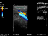
What is duplex scanning?
The best modern diagnostic method
is ultrasonic scanning, since the quality of the results obtained is maximum. In the West, preference is given to color mapping. It increases the accuracy of the information if you need to track the speed of blood flow towards and away from the sensor (marking is done in red and blue). The method allows you to perform the following tasks:
- Monitor damage and deformation of the venous walls.
- Find out the speed of blood flow in the deep and superficial veins.
- Check the functionality of the valves.
- Assess the dimensions and density of the blood clot (determine the degree of thrombosis).
Classic and minimally invasive surgeries are always performed with 100% accuracy, as they do ultrasound of the veins of the lower extremities in combination with duplex scanning.
How is the procedure performed?
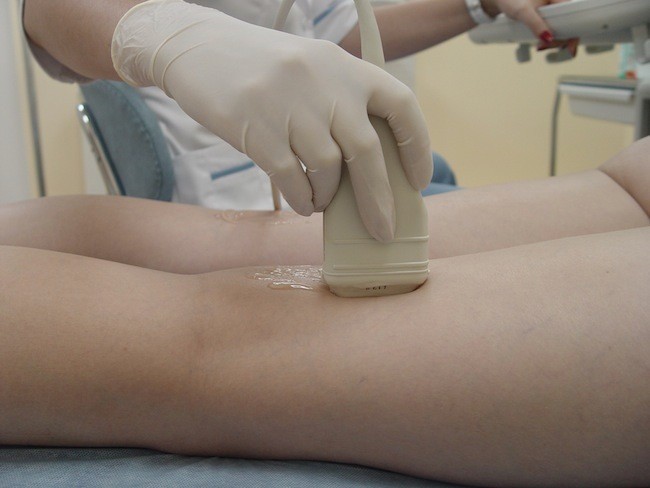
Before visiting a specialist, be sure to take a shower . There is no need to shave your leg hair or deep clean your skin. It is advisable not to drink alcohol on the eve of an ultrasound examination. The procedure is performed as follows:
- In the office you undress from the waist down (down to your underwear).
- Ultrasound gel is then applied to the legs.
- The specialist checks the condition of the veins, valves, vessels and arteries using a device with a sensor.
- If necessary, the intensity of pressure and the power of the beam are increased to more clearly see the condition of the deep veins.
- Your body position changes—your doctor may ask you to stand up or lie down to see how the blood flows. For getting additional information you will need to hold your breath (take a deep breath).
Now you know exactly how they do it . The session lasts a few minutes and you will not experience any discomfort. Painful sensations are missing. During the change in ultrasound intensity, you will not feel anything (many people fear pain or discomfort).
Do not forget that Only a competent phlebologist can decipher the result with great experience. Therefore, immediately after a duplex scan or ultrasound, go to an appointment with a specialist so that he can make a final conclusion.
The First Phlebology Center will determine the condition of the veins of the lower extremities and prescribe an effective course of treatment for you. You can sign up for an examination right now by phone.
We practice following methods Vein disease research:
- Duplex scanning of the veins of the lower extremities has already become the gold standard in the diagnosis of venous diseases.
Ultrasound of the veins of the lower extremities is performed to assess the condition of valves, veins and blood vessels. During the examination, the doctor pays attention to all changes - neoplasms (blood clots) or low speed blood flow Ultrasound examination is relevant for the reason that this procedure identifies a specific area in which pathological processes appear. To perform the diagnosis, no complex preparatory process is required.
Send a request and we will contact you
with you soon
Fact: A surgeon or phlebologist should evaluate the diagnostic result after ultrasound. Only a professional can determine the pathology of the veins and accurately decipher the result of an ultrasound examination.
Which diagnostic methods accurately reflect the result?
The main method is to reflect the beam through red blood cells, which allows 100% determination of the condition of the veins and arteries. The method demonstrates the speed of blood flow and all pathologies, therefore it is deservedly popular all over the world.
Nowadays, Doppler ultrasound (abbreviated as Doppler ultrasound) is used. Ultrasound ultrasound is used to check the patency of the veins and the condition of the valves. The method is highly accurate.
Duplex scanning is one of the methods that is used the best clinics USA and Europe. Thanks to it, all defects in the functioning of the venous system are revealed. It has much in common with ultrasound of the veins of the lower extremities using the Doppler ultrasound method. During the diagnostic process, the following is revealed:
- Valve condition.
- Presence of blood clots.
- Thrombosis degree.
- Patency of superficial and deep veins.
- Condition of the venous walls.
Color mapping is often used, which makes it possible to estimate blood flow speed with maximum accuracy. blue tint shows movement from the sensor, and red oncoming traffic. Speed directly depends on intensity.
In what cases is it worth performing an ultrasound of the veins?
A specialist will recommend you undergo this procedure during your primary medical examination. The procedure may be prescribed for the following alarming symptoms: 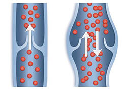
- Dilatation of veins.
- Swelling.
- Trophic ulcers.
- Change in color of the epidermis.
- Night cramps.
- Burning sensation for no apparent reason.
Diabetics, smokers, athletes and genetically predisposed people are at risk. Among the symptoms of vein diseases, there are such rare phenomena as muscle weakness throughout the day, pain when running or walking, and pale skin.
How is ultrasound performed?
To carry out this procedure, you do not need to follow a diet or prepare for several days; you just need to take a shower before visiting medical center. The procedure is very simple and does not require any effort from you:
- You undress from the waist down to your underwear.
- Gel is applied to the feet.
- The doctor runs the sensor along the leg.
- The ultrasound intensity changes periodically.
A change in intensity is required to check the deep veins and blood vessels. This does not affect your feelings in any way and does not lead to negative consequences.
When using the Doppler ultrasound method, the procedure becomes somewhat more complicated. The doctor applies cuffs to assess your blood pressure. The examination is carried out both standing and lying down.
Maximum accuracy guaranteed
The First Phlebological Center has imported equipment and performs the ultrasound procedure with maximum accuracy. This allows for a correct diagnosis, which will lead to timely treatment diseases. You can make an appointment by phone during business hours.
When scanning in B-mode, intact veins have a thin, elastic wall, a homogeneous and echo-negative lumen, completely compressed by the ultrasound sensor. In a lying position, their diameter is elliptical or disc-shaped. IN vertical position the diameter of the vein increases (by an average of 37%), it acquires a rounded shape (Fig. 1).
Rice. 1. Vascular bundle of the popliteal fossa (intact popliteal vein - PCV).
Also, normally, a noticeable movement of blood can be recorded in the lumen of the vein, that is, the movement of the flow of blood particles is visualized in the form of whitish point echoes moving in accordance with the breathing cycles.
Indicators of normal diameter of venous vessels are presented in tables 1, 2.
A distinctive feature of the venous system is the presence of valves. Valves are typically bicuspid folds of endothelium concave toward the heart that allow blood to flow in one direction. The valves are often quite clearly visible, mainly in the lumen of large veins, and are identified in the lumen of the vein at different levels limbs. The leaflets of a functional valve are attached to the wall of the vein with one edge, and freely oscillate in its lumen with the other. The movements of the valves are synchronized with the phases of breathing. On inhalation they are in a parietal position, on exhalation they converge in the center of the vessel (Fig. 2). In this way, blood is emptied from the valvular sinuses. Usually the valve looks like two thin, highly echogenic, whitish, no more than 0.9 mm thick, bright stripes in the lumen of the vein. However, very often the valve leaflets may not be clearly depicted, but only outlined by the echogenicity of the blood flow around them. This effect is the result of an increase in blood density and blood stagnation, which tends to form in the area of the valvular sinuses (the effect of “smoke” and valve “nest”) (Fig. 3). The ability to enlarge the image allows you to clearly record the valve leaflets, observe their “flight” in the blood flow and “slamming” at the height of hydrodynamic loads.
Rice. 2. Normal valve in the superficial femoral vein.
Rice. 3. Popliteal vein valve in B-mode. Hypoechoic signals from blood particles are detected in the lumen of the vein and valvular sinuses).
Small tributaries, ranging from 1 to 3, are often drained into the area of the valvular sinuses. More often there is a single valveless inflow with a diameter of 2-3 mm, flowing into the projections of the valvular sinus at different levels. In the valves of the brachial veins, tributaries are detected in 78.2% of cases, in the area of the permanent valve of the superficial femoral vein, which is located immediately under the mouth deep vein hips, 1 or 2 similar tributaries can be found in 28.3% of the limbs. A high frequency of sinus tributaries is noted in the valves of the popliteal vein, with 2 tributaries (the mouths of which were located in both sinuses) in 50.4% of cases, 1 tributary in 41.8%, 3 tributaries in 1.8%. Their distinctive feature was the presence of monocuspid estuarine valves.
The physiological feasibility of equipping venous valves with tributaries is explained by the fact that the flow of blood from the muscular tributaries into the sinuses of the valve, along with retrograde blood flow, causing the closure of the valve leaflets, prevents the process of thrombus formation due to the washing out of the formed elements of blood from the sinuses. The location of the orifices of the tributaries in the projection of the valvular sinus and the direction of the stream of incoming blood can change the position of the valve leaflets, which is rational for their closure. It is not excluded possible role valveless inflows in damping supravalvular hypertension under the influence of retrograde blood flow. The listed mechanisms to a certain extent contribute to the normal function of the venous valve, however, they sometimes cause eccentric venous reflux, leading to valvular incompetence. The constancy of the location of the tributaries in the valves of the popliteal vein, which bear the highest hemodynamic load, also indicates their functional significance.
When performing hydrodynamic tests that cause a wave of retrograde blood flow (Valsalva maneuver, proximal compression of the muscle mass), the valve leaflets close tightly and are visualized either directly in the form of an echogenic line, or indirectly in the form of a contour image formed as a result of an increase in the echo density of blood in the supravalvular zone, caused by her temporary stasis. In this case, the line of closure of the valve flaps is clearly recorded when scanning in M-mode. The Dopplerogram shows a short wave of retrograde blood flow. Its duration is 0.34±0.11 seconds. The lumen of the vein in the area of the valvular sinus expands in a balloon-like manner. The Dopplerogram returns to the isoline, again intensifying upon exhalation or removal of compression. In quiet orthostasis, the valves of the main veins (femoral, popliteal) are constantly open, their valves are at an angle of 20-30° relative to the vein wall. The valve leaflets perform a floating flight in the lumen of the vein with a high frequency and small amplitude - 5-15o. The closure of the valve leaflets, both in clino- and orthostasis, occurs only with forced breathing or imitation physical activity associated with abdominal wall tension. When simulating walking with the involvement of the muscle mass of the lower leg and thigh, the valve valves are constantly open, only a significant increase in linear and volumetric velocities is noted on the Dopplerogram.
The functionality of valve structures is also studied in the modes of color circulation and power Doppler. By encoding the movement of blood particles between the venous wall and the valve leaflet, color flows provide an indirect idea of the shape of the valve and the state of its leaflets. Normally, when breathing, blood flow in a vein is mapped (coded) in one color. During take a deep breath blood flow is not recorded, and the lumen of the vessel becomes echo-negative.
Table 1. Indicators of the diameter of the venous vessels of the femoral segment
Table 2. Indicators of the diameter of the venous vessels of the calf segment
IN horizontal position color mapping of the main veins determines laminar flow blood with a specific color code (Fig. 4). Pulse Dopplerography records a unidirectional phase flow that coincides with the subject’s breathing, decreasing with inhalation and increasing with exhalation, which is a reflection of the predominant influence of the vis a frontе phenomenon (a set of factors that determine blood suction) on venous drainage in a supine position (Fig. 5).
Rice. 4. Antegrade blood flow in the lower third of the superficial femoral vein in the color flow mode.
Rice. 5. Spectral profile of normal venous blood flow.
Each large Doppler wave in large-caliber veins is split into smaller waves, the frequency of which coincides with the heart rate, which characterizes such a venous return factor as the suction action of the heart, which is one of the components of the vis a frontе factor. The fact that these waves belong to the activity of the heart chambers (right atrium), and not to the transmitting pulsation of the artery accompanying the vein, is evidenced by the fact that this phenomenon is also present when examining veins in patients with occlusive lesions of the corresponding arterial segment.
When the subject holds his breath while exhaling, the Dopplerogram acquires a low-amplitude continuous-wave character with peaks corresponding to the heart rate. This test allows you to evaluate the second factor of venous return - the vis a tergo factor (residual cardiac output). The influence of these forces of venous return is interrelated, one of them (vis a tergo) provides a pushing effect, the other (vis a frontе) provides a suction effect. There is no doubt that the tone of the tissues surrounding the vein is also important for the implementation of the listed return factors.
It should be noted that the speed of blood flow in the main veins increases from the periphery to the center. In a standing position, the blood flow rate decreases significantly (by an average of 75%). The Dopplerogram takes on a discrete wave form, synchronized with the act of breathing, while the respiratory waves have a more distinct phase than in the supine position. At the height of inspiration, the Dopplerogram curve comes to the isoline. To exclude the influence of respiratory movements on venous return, the subject holds his breath as he exhales. In this case, the Dopplerogram curve takes on a characteristic discrete wave form with a wave frequency that coincides with the heart rate. The appearance of discreteness indicates that the vis a tergo factor is leveled by the orthostatic position. Thus, in a standing position at rest, the venous return is mainly influenced by the vis a fronte factor.
Indicators of antegrade venous blood flow in horizontal and vertical positions are presented in Table 3.
Table 3
Indicators of antegrade blood flow in healthy individuals
Note. Vmean, — average linear speed; Vvol ~ volumetric velocity; OBV - general femoral vein, GSV - large saphenous vein, PCV - popliteal vein;
Also during ultrasound examination is carried out quantification indicators of phlebohemodynamics (regional).
Table 4 shows normal indicators antegrade venous blood flow: maximum linear velocity in the spectrum; time-averaged value maximum speeds in the spectrum; volumetric blood flow velocity.
The parameters of the retrograde blood flow wave that occur during hydrodynamic tests (Valsalva maneuver, compression (cuff) test) are also assessed: duration of reflux; linear velocity of retrograde blood flow; acceleration of reflux.
Table 4. Quantitative indicators phlebohemodynamics in practically healthy individuals
|
Options* |
Anatomical localization of the venous vessel | ||||||
| OBB | GSV | PBB | WBG | PV | MPV | ZBV | |
| Velocity indicators of antegrade blood flow: | 13.9±2.1 7.85±0.2 |
12.6±1.8 5.7±0.5 |
11.9±1.4 4.9±0.4 |
11.8±1.8 3.8±0.3 |
14.2±1.9 7.2±0.4 |
7.2±1.1 1.0±0.3 |
4.8±1.2 0.4±0.1 |
| Indicators of induced retrograde blood flow: | ≤0,5 | ≤0,5 | ≤0,5 | ≤0,5 | ≤0,5 | ≤0,5 | ≤0,5 |
*Note: Vm – maximum linear velocity in the spectrum, cm/sec;
TAMX – averaged linear velocity in the spectrum, cm/sec;
Vvl – volumetric blood flow velocity, ml/sec;
T – duration of reflux, sec;
Vr – linear velocity of retrograde blood flow, cm/sec;
Accl – acceleration of reflux, cm/sec2.
- ← Chapter 4. Diagnosis of disorders of blood outflow from the lower extremities.
- Content
- → 4.2. Duplex scanning for varicose veins.
What worries you?
Ultrasound of the vessels of the extremities is one of the most modern, safe and informative ways to study the condition of the vessels of the arms and legs. An ultrasound wave is able to show vessels in a graphical form, which allows you to diagnose various vascular diseases. In this article we will talk about how this procedure is carried out, whether it requires special preliminary preparation, for what indications it is prescribed and what diseases it can diagnose.
How is ultrasound of the vessels of the arms and legs performed?
Analysis of the properties of blood flow in the extremities is possible due to the ability of ultrasound waves to reflect from those formed blood particles that are in motion. This type of imaging is called Doppler, also known as Doppler ultrasonography. The research procedure looks something like this.
Before scanning begins, the doctor applies to the surface of the skin being examined. a large number of a special gel that will eliminate the air gap between the sensor and the human body. If such a gel is not used, the signal will be much weaker, and the image on the screen will be unclear and incomprehensible.
- First, the doctor examines the vessels in a lying position, with the limb in a bent state.
- The next stage: if the procedure is performed on the leg, then the patient is asked to take a vertical position, if on the arm, then it will be enough to straighten it.
- For each patient, the physician manually selects a different frequency of the ultrasound wave; it can vary depending on how deep the vessels are located.
- The frequency of ultrasound also depends on how deep the detail is required; most often the frequency ranges from 6 to 12 megahertz.
- Very deep and small veins are examined using a special low-frequency sensor.
Preparation for the procedure
This type of scanning does not require special preparation from the patient; he can visit the ultrasound room any day. Typically, a referral is written by the attending physician; he provides the diagnostician with the necessary amount of information to conduct a detailed study. Most often, a preliminary diagnosis and those areas that need more attention are also indicated. close attention. The only thing a person should do before an ultrasound scan of the vessels of the lower extremities, as well as the upper ones, is to choose comfortable clothes that will allow you to easily open the required area of skin on the arm or leg. When researching upper limbs It is recommended to remove all jewelry. You should bring a diaper and wipes with you to your appointment, which can be used to remove any remaining gel from the skin.
When is an ultrasound of the vessels of the arms or legs prescribed?
The decision on the need for such a procedure is made by the attending physician, this may be a traumatologist or. Indications for ultrasound of the vessels of the lower extremities, as well as the hands, will be:
- V initial stage or in a chronic form.
- , especially if it is accompanied by headaches and deterioration in general health.
- If walking causes pain or the patient experiences discomfort when moving the upper limbs.
- In cases where the patient complains of night pain in the joints of the upper limbs or legs. This is especially true for older people, in cases where the pain subsides when lowering their legs or arms from the bed.
- With severe excess body weight, obesity.
- If the patient smokes a lot and for a long time long period time.
- If you have had surgery on the upper or lower extremities.
- In cases where a person has increased content cholesterol in the blood.
Often, the indication for such a procedure may not be the doctor’s suspicions about a specific disease, but the patient’s complaints. Ultrasound of the vessels of the lower extremities is prescribed when the following alarming symptoms appear:
- The legs swell a lot, especially in the afternoon.
- There is a strong effect that will be noticed not only by the doctor.
- At night, your legs cramp or cramp.
- In cases where dilated veins appear in the legs during pregnancy, the legs ache and hurt.
- There are any types acute pain accompanied by an increase in body temperature.
- The skin on my legs suddenly changed color.
- There are trophic ulcers on the limbs.
Interpretation of the results of ultrasound examination of the extremities
If you had a scan in a regular ultrasound room, you will be given a conclusion about the condition of your blood vessels, which you will not be able to figure out on your own. In this case, you need to contact your doctor for a transcript. He will not only evaluate the ultrasound indicators, but also correlate them with the examination data and laboratory research. Only after this will you be given a final diagnosis and appropriate treatment will be prescribed. In cases where an ultrasound of the vessels of the lower extremities or arms will be performed directly by a vascular surgeon or doctor, the scanning results will be interpreted directly after the procedure and a diagnosis will be made immediately. The assessment of vascular blood flow is carried out using the following indicators:
- The highest blood flow velocity recorded during systole.
- The minimum blood flow velocity that is recorded in diastole.
- Vascular resistance.
- Pulsation index.
- Thickness of vessel shells: inner and middle.
Contraindications to ultrasound of the extremities
There are many versions of doctors about the dangers of ultrasound, but none of them have been proven. Therefore, we can say with confidence that this method is safe, ultrasonography upper limbs and legs, if there is appropriate doctor's indications, can be performed even on children and pregnant women. But you should refuse the procedure if the patient has:
- Burns on the hands or feet.
- The body develops infectious diseases or inflammatory processes.
- There are skin diseases in the form of rashes or irritations.
- There are wounds or ulcers at the intended examination site.
- Sudden attacks or bleeding.
What diseases can ultrasound of the limbs and their vessels detect?
If immediately upon occurrence alarming symptoms If you conduct an ultrasound of the lower extremities or arms, it will be much easier to cure the disease. Using ultrasound scanning of the vessels of the legs and arms, the following diseases can be identified:
- , is characterized by a disruption of normal blood flow, while the legs or arms hurt and ache.
- Endarteritis is diseased small vessels, characterized by the appearance of goosebumps on the skin of the extremities.
- Phlebeurysm.
Ultrasound of the veins of the lower extremities is a procedure for studying the anatomy of these vessels, the condition of their valves, the characteristics of blood flow in them, based on. Such a study helps not only to determine in which area and due to which veins blood circulation is affected, but also to identify blood clots and inflammation. The method is non-invasive and safe and can be performed frequently without requiring any preparation.
The results are interpreted by a phlebologist or vascular surgeon. At the end of the article you can see educational video with doctor's comments this species examinations.
Types and purposes of ultrasound of the veins of the lower extremities
The method is based on the Doppler effect, that is, the reflection of an ultrasound beam from red blood cells moving in blood vessels. This gives the doctor information about how the vascular cord passes, how high the speed of blood flow is in it, and clarifies other nuances that differ when examining arteries and veins.
Based on this, ultrasound diagnostics of veins and resistance vessels (arteries) of the lower extremities is also called “Doppler”. It has several subspecies.
What is ultrasound
This is the most “ordinary” Doppler ultrasound. It is useful for:
- determining the patency of deep venous collectors
- assessing the patency of superficial venous vessels
- diagnostics of the condition of venous valves
- assessment of the condition of the valves of the perforating veins only if they are located in a typical manner (otherwise such ultrasound will not see them).
Duplex scanning (USDS)
This method is the most common and most accurate in assessing the venous system. It is a combination of ultrasound and online scanning. This ultrasound examination of the venous collectors of the lower extremities has the following goals:
- determine the condition of the venous walls
- assess the condition of the valves of any veins
- analyze the patency of deep and superficial venous collectors
- assess the condition of the perforating veins, wherever they are located
- see a blood clot, determine its size, location, mobility
- determine the degree of venous thrombosis.
Ultrasound with color mapping
This is the most modern method Ultrasound examination of the vessels of the extremities. Here, in addition to assessing the characteristics of blood flow in real time, there is also a color indication of blood flow speed. All shades of red will indicate blood flow going towards the sensor, shades of blue – directed away from it. The more intense the hue, the higher the speed.
Now this method is beginning to take an increasingly leading position in diagnosing problems of the arterial and venous vessels of the body.
Who needs to undergo Doppler ultrasound of the extremities?
Performing an ultrasound scan of the veins of the lower extremities is urgent for the following symptoms:
- swelling of the legs
- dilated veins visible to the eye
- change in the color of the leg (usually these are areas Brown or a large section of purple-pink color)
- trophic ulcers on the leg
- frequent cramps in the leg muscles
- numbness, tingling of the legs
- itching in the legs in the absence of skin diseases
Doppler ultrasound scanning of the leg arteries is indicated if:
- you suffer from diabetes
- your blood cholesterol levels are high
- Do you smoke
- if you have pain in your legs that gets worse when walking
- pale lower limbs
- your feet get cold quickly, even if only your feet get cold
- leg muscle weakness
- a feeling of “goosebumps” on the skin of the legs.

Ultrasound diagnostics of the veins of the upper extremities has its own indications:
- numbness of hands
- weakness in the hands
- hands or arms become completely cold quickly
- Wounds on the upper extremities do not heal for a long time
- especially if these symptoms appear in smokers, hypertensive patients, people with diabetes
- if blood pressure measured at different hands, has a difference of 20 mm Hg or more.
Preparing for the study
Preparation for angioscanning of the venous vessels of the extremities is not necessary. You just need to carry out the usual routine hygiene measures (by the way, you can do without such preparation).
Physiology of venous vessels in the legs
There are as many as three venous systems in the lower extremities, united into a single network. So, there are deep and superficial venous collectors, they have a large number of bicuspid valves that help blood move against gravity - from bottom to top.
The main venous vessels are deep, they are not visible to the eye even with thin skin and a small number subcutaneous tissue. Almost 90% of the blood flows through them; their wall has a small number of muscle fibers (unlike superficial veins).
Deep and superficial venous system communicate via communicating (perforating) veins, of which there are about 100 in each leg. In them, blood moves from bottom to top, from superficial to deep collectors. This movement is helped by pressure in the right atrium, as well as by the work of muscles that necessarily contract and relax in an upright position of the body.
How is the examination done?
An ultrasound scan of the legs looks like this.
- You come to the office, undress from the waist down, leaving your underwear on.
- Next, a little cool gel is applied to your leg - first one, then the other. They will move a sensor along it.
- Depending on the depth of the vessel being examined, the doctor will change the frequency of the emitted ultrasound, but this is not felt at all.
Ultrasound scanning of the arterial vessels of the legs has the peculiarities that during the procedure different areas limbs (and upper ones too), the doctor will apply cuffs and measure blood pressure. The examination will be carried out standing and lying down.
How to do an ultrasound diagnosis of leg veins: first, these organs are examined while lying down, then while standing. Tests are also carried out to determine how blood will move from superficial to deep vessels. To do this, ask to take a deep breath, then, without exhaling air, strain.
Ultrasound scanning of the deep venous collectors of the lower extremities is performed in color and normal modes. In order to better visualize these veins, not only straining tests are performed, but also pressure is applied to the area where they are located with different forces.
Ultrasound of the vessels of the lower extremities is performed: standing, lying on the stomach, lying on the back.
How to decrypt data
1. Examination of the arteries of the legs
Ultrasound of the leg arteries assesses:
- vessel anatomy
- thickness vascular wall(may be written “intima-media complex”)
- indicators of vascular resistance (they have the same names that are used for), on the basis of which the patency and narrowing of the arteries are judged
- characteristics of blood flow depending on the phase of the heart (systole or diastole)
- the speed of blood flow, the dynamics of its change depending on which vessel is examined.
Thus it is described:
- type of blood flow in each artery (there are norms for each of them)
- peak velocity for each artery (there are also normative tables for them)
- pulsation index (PI) - the ratio of the total sum of peak speeds to the average speed: should be greater than 4 on the legs
- damping factor (DF), that is, the ratio of the pulsation index measured on the far parts of the limb to PI on those closer to the heart. Its norms are 1.15-1.48 (artery stenosis - if DF is less than 1)
- meaning blood pressure on each artery: there are norms for arteries, plus the difference for the same artery on different legs(or hands) should not be more than 20 mm Hg
- The malleolar-brachial index (BBI), that is, the ratio of blood pressures at the ankle and shoulder, should be about 1.0
- resistive index (RI), which is measured using ultrasound on each artery (there are standards for them). This is the ratio of the difference between the maximum and minimum speeds in the vessel to the maximum speed
- state of the intima-media complex femoral artery(its normal thickness is 1.0-1.2)
- the percentage of artery stenosis is measured
- if there are plaques, the following is described: localization, degree of their mobility (mobile or not), degree of homogeneity (consist of one substance or not), whether they are complicated or not, and what they are complicated by.
2. Vein examination
It is carried out according to the ultrasound protocol of the venous vessels of the legs:
- Each vein on the right and left is assessed
- it is noted whether the blood flow in the veins is related to breathing
- describes whether these collector vessels are well compressed by the sensor
- is there any thickening of the wall
- are there additional masses (thrombi) in the lumen?
- Are the vein valves healthy?
- Are there pathological refluxes (reflux) through the veins?
- describe exactly where incompetent perforating veins with a diameter of 3 mm or more are located.
If there is a thrombus, ultrasound of the deep venous collectors describes its characteristics:
- by what percentage does it block the lumen of the vein (if it does)
- if the thrombus does not narrow the lumen, it is described as parietal or floating (mobile)
- soft or dense
- whether the thrombus moves when touched by the sensor or not.
Based on all this, a conclusion is drawn, based on which, only a phlebologist can determine further treatment tactics.
Where to take the study
It is best to find out from a vascular surgeon where to do an ultrasound diagnosis of the veins of the lower extremities. He can do it himself in a multidisciplinary center or specialized clinic, can recommend you a specialist whom he trusts.
You can do it for free at the branch vascular surgery large public hospital.
You can undergo a test for a fee at your nearest phlebology clinic or multidisciplinary center, where you can ask by phone how much an ultrasound scan of the venous section of the lower extremities costs. In the offices ultrasound diagnostics where all organs are examined, it is better not to undergo such a procedure.
The price of ultrasound angioscanning of veins depends on which vessels you want to look at, so to speak, “what Doppler” you need to undergo and what type of it.
For example, the price for duplex ultrasound of the arteries of the legs ranges from 1300 to 3500 rubles. The price of ultrasound diagnostics of the veins of the extremities depends on the type of diagnosis. Thus, duplex angioscanning costs from 800 rubles to 5 thousand rubles, color vascular duplex scanning– from 800 to 6500 rubles.
The average price for ultrasound of the venous and arterial sections of the lower extremities is about 2-2.5 thousand rubles.
Thus, ultrasound of the veins of the lower extremities is a safe and painless diagnosis that can be performed on anyone at any age. Having completed it without any preparation, you will already know in an hour how things are going with your blood vessels (this is especially important for those who have several risk factors for vascular diseases).

ATTENTION! The information on the site is for reference or popular information only. Correct treatment and purpose medicines can only be carried out by a qualified specialist, taking into account the diagnosis and medical history.
Successful diagnosis and treatment, health and feeling great!.
UziLab