Organs of the thoracic cavity: structure, functions and features. Anatomy of the human chest Chest changes
Rib cage
the set of thoracic vertebrae of the thoracic ribs and sternum (Sternum), which in reptiles, birds, mammals and humans provides a strong support for the shoulder girdle and allows the use of intercostal muscles during respiratory movements. Historically, G. appears in amniotes (See Amniotes) in connection with the progressive development of their organs of movement and breathing. In mammals, the respiratory function of the gastrointestinal tract is enhanced due to the appearance of a thoraco-abdominal obstruction (See Thoracic obstruction) and the formation of the thoracic cavity (See Thoracic cavity). In most reptiles, whose body touches the ground, the abdominal cavity is flattened from top to bottom and its lateral diameter is greater than the dorsal diameter; In mammals and some reptiles (for example, chameleons), in which the body is raised on its paws from the ground, the abdominal cavity is flattened laterally and its dorsal diameter predominates over the lateral one. This form of G. to. is called “primary”. In apes and especially in humans, the primary form of gastrointestinal tract changes to a “secondary” one, in which the lateral diameter exceeds the spinal diameter. A barrel-shaped blood vessel with equal dorsal and lateral diameters is characteristic of animals that gallop on hind legs(kangaroos, jerboas), flying (birds, the bats, from fossils - pterosaurs), swimming (whales, from fossils - ichthyosaurs). G. in humans has the shape of flattened in the anteroposterior direction truncated cone. There are lateral walls of the rib cage, which are formed by 12 pairs of ribs separated intercostal spaces; the anterior wall, which includes the ends of the ribs and the sternum, and back wall with a spine in the middle. On top of the chest there is an opening - the upper aperture, the boundaries of which are the right and left first ribs, the first thoracic vertebra and the manubrium of the sternum. The trachea, esophagus, vessels and nerves pass through this opening into the chest cavity. The lower aperture is limited by the ends of the ribs. Below, the G. k. is separated from abdominal cavity diaphragm. Depending on gender, age, body type, there are various shapes G. k., for example, in men the G. k. is more cone-shaped, in women it is cylindrical. Children suffering from rickets are distinguished by a keeled hemorrhage; in the elderly, the hemorrhoid is either flattened or becomes barrel-shaped, especially with pulmonary emphysema (See Pulmonary emphysema). Persons of an asthenic physique (see Human Constitution) have an elongated and flattened body; in people of the picnic type, the body is short and massive. When you inhale, the blood vessel expands, which is accompanied by an increase in its longitudinal, anteroposterior and transverse dimensions. V. V. Kupriyanov.
Great Soviet Encyclopedia. - M.: Soviet Encyclopedia. 1969-1978 .
Synonyms:See what “Chest” is in other dictionaries:
Rib cage- (compages thoracis) consists of ribs connected at the anterior ends to the sternum (sternum), and at the posterior ends to the thoracic vertebrae. The frontal surface of the chest, represented by the sternum and the anterior ends of the ribs, is significantly shorter than... ... Atlas of Human Anatomy
RIB CAGE- (thorax), composed of the thoracic spine at the back, twelve pairs of ribs and their cartilages at the sides and the sternum at the front. Usually only the first seven pairs of ribs, rarely eight, reach the sternum; VIII, IX and usually X ribs are connected with their cartilages to... ... Great Medical Encyclopedia
The combination of thoracic vertebrae, ribs and sternum, which forms a strong support for the shoulder girdle in reptiles, birds, mammals and humans. The space inside the chest (thoracic cavity) in mammals is separated from the abdominal... ... Big encyclopedic Dictionary
- (thorax), in anatomy, the part of the body between the neck and the abdominal cavity. In mammals, it is formed by the rib cage and contains the lungs, heart and esophagus. Separated from the abdominal cavity by the DIAPHRAGM. In arthropods it consists of several segments, to which ... Scientific and technical encyclopedic dictionary
- (thorax), part of the axial skeleton of amniotes, formed by the connection of the thoracic vertebrae, thoracic ribs and sternum into a single system. It arose for the first time in reptiles in connection with the progressive development of the organs of movement (support of the shoulder girdle) and breathing... Biological encyclopedic dictionary
Noun, number of synonyms: 1 breast (33) Dictionary of synonyms ASIS. V.N. Trishin. 2013… Synonym dictionary
Bones of the human chest The chest, chest (lat. Thorax) is one of the parts of the body. Formed by the sternum, ribs, spine... Wikipedia
The combination of thoracic vertebrae, ribs and sternum, which forms a strong support for the shoulder girdle in reptiles, birds, mammals and humans. The space inside the chest (thoracic cavity) in mammals is separated from the abdominal... ... encyclopedic Dictionary
RIB CAGE- chest, skeleton thoracic bodies of vertebrates. Consists of osteochondral segments, each of which includes a vertebra, a pair of ribs, and a fragment of the breastbone (sternum). At the large cattle 13 x 14 segments, y... ... Veterinary encyclopedic dictionary
- (box, thorax) has a barrel shape in humans different shapes and is made up of bones: 12 pairs of ribs, 12 thoracic vertebrae and sternum. The posterior ends of the ribs are attached to the vertebrae by means of ligaments; anterior at the upper 7 ribs (true ribs)… … Encyclopedic Dictionary F.A. Brockhaus and I.A. Ephron
Books
- Radiation diagnostics. Chest, M. Galanski, Z. Dettmer, M. Keberle, J. P. Oferk, K. I. Ringe, The book is part of the "Dx-Dircct" series, dedicated to imaging methods for diagnosing various organs and systems. All books in the series are built according to a single scheme, which provides an overview... Category: Ultrasound. ECG. Tomography. X-ray Series: Dx-Direct Publisher: MEDpress-inform,
- Radiation diagnostics Chest, Galanski M., Dettmer Z., Keberle M., Oferk J., Ringe K., The book is part of the “Dx-Direct” series, dedicated to imaging methods for diagnosing various organs and systems. All books in the series are built according to a single scheme, which provides an overview... Category:
The chest (thorax) (Fig. 112) is formed by 12 pairs of ribs, sternum, cartilage and ligamentous apparatus for articulation with the sternum and 12 thoracic vertebrae. All these formations form the chest, which in various age periods has its own structural features. The chest is flattened from front to back and expanded in the transverse direction. This feature is affected vertical position person. As a result internal organs(heart, lungs, thymus gland, esophagus, etc.) exert pressure primarily not on the sternum, but on the diaphragm. In addition, the shape of the chest is influenced by the muscles that move the shoulder girdle, starting on the ventral and dorsal surfaces of the chest. The muscles form two muscle loops that exert pressure on the chest from front to back.
112. Human chest (front view).
1 - apertura thoracis superior;
2 - angulus infrasternalis;
3 - apertura thoracis inferior;
4 - arcus costalis;
5 - processus xiphoideus;
6 - corpus sterni;
7 - manubrium sterni.

113. Schematic representation of the shape of the chest of a person (A) and an animal (B), (according to Benninghoff).
In animals, the chest is compressed in the frontal plane and extended in the anteroposterior direction (Fig. 113).
The first rib, the manubrium of the sternum and the first thoracic vertebra limit the upper aperture of the chest (apertura thoracis superior), which has a size of 5x10 cm. The boundaries of the lower aperture of the chest (apertura thoracis inferior) are the xiphoid process of the sternum, the cartilaginous arch, the XII vertebra and the last rib. The size of the lower hole is significantly larger than the upper one - 13x20 cm. The circumference of the chest at the level of the VIII rib corresponds to 80 - 87 cm. Normally, the latter size should not be less than half of a person’s height, which characterizes the degree of physical development.
Through the upper aperture of the chest, the trachea, esophagus, large blood vessels and lymphatic vessels, nerves. The lower aperture is closed by a diaphragm through which the esophagus, aorta, and lower vena cava, thoracic duct, vegetative trunks nervous system and other vessels and nerves. The intercostal spaces, in addition to ligaments, are filled with intercostal muscles, vessels and nerves.
During inhalation and exhalation, the size of the chest changes.
This is only possible due to the large length and spiral structure of the ribs. The posterior end of the rib is fixed to the spine by two joints (the head of the rib with the vertebral body, the tubercle of the rib with the transverse process), located on the same bone and motionless in relation to each other. Therefore, the movement occurs in both joints simultaneously, namely: rotation of the back of the rib along the axis connecting the joint of the head of the tubercle of the rib. Anatomically, these joints have a spherical shape, but functionally they combine and form a cylindrical joint (Fig. 114). When the posterior end of the rib rotates, its anterior spiral part rises up, moves to the sides and anteriorly; Due to this movement of the ribs, the volume of the chest increases.

114. Scheme of movement of the ribs.
A - location of the rotation axes of individual ribs.
B - rotation diagram of the I and IX ribs (according to V.P. Vorobyov).
Age characteristics . In a newborn, the chest resembles in shape the chest of animals, in which, as is known, the sagittal size predominates over the frontal. In a newborn, the heads of the ribs and their anterior ends are almost at the same level. At 7 years of age top edge The sternum corresponds to level II - III, and in an adult - III - IV thoracic vertebrae. This lowering is associated with the appearance of thoracic breathing and the formation of a spiral-shaped ribs. In cases where rickets disrupts mineral metabolism and delays the deposition of salts in the bones, the chest takes on a keeled shape - “chicken breast”.
The substernal angle in a newborn reaches 45°, after a year - 60°, at 5 years - 30°, at 15 years - 20°, in an adult - 15°. Only from the age of 15 are gender differences in the structure of the chest noted. In men, the chest is not only larger, but there is a steeper bend of the rib in the corner area, but the spiral twisting of the ribs is less pronounced. This feature also affects the shape of the chest and the nature of breathing. Due to the fact that in women, as a result of the pronounced spiral shape of the ribs, the anterior end is lower, the shape of the chest is flatter. Therefore, in women, the thoracic type of breathing predominates, in contrast to men, who breathe mainly due to the displacement of the diaphragm (abdominal breathing).
It has been noticed that people of different builds also have their own chest shape. People of short stature with a voluminous abdominal cavity have a wide but short chest with a wide lower opening. On the contrary, tall people have a long and flat chest shape.
In the elderly, the elasticity of the costal cartilages significantly decreases, which also reduces the excursion of the ribs during breathing. In old age, due to frequent breast cancer, the shape of the chest also changes. Thus, with emphysema, a barrel-shaped chest is often observed.
Physical exercise has a significant shaping effect on the shape of the chest. They not only strengthen the muscles, but also increase the range of motion in the joints of the ribs, which leads to an increase in the volume of the chest and the vital capacity of the lungs when inhaling.
The chest is a frame consisting of a set of bones and separated from the abdominal cavity by a flat respiratory diaphragm. Due to its structure of a closed hollow space, this part of the body protects the internal organs from mechanical influences from the environment.
Thorax skeleton
The skeleton of the human chest includes:
- ribs
- sternum.
Thoracic vertebrae
They are 12 unpaired bones, each of which is a supporting unit of the spine and has a massive anterior fragment - the vertebral body. The body is designed to take on the main load and, together with the arch, forms a ring within which the spinal cord is located. The vertebrae are connected to each other by discs and a whole network of ligaments and muscles that ensure the flexibility of the column.
An adult's discs together can account for a quarter of the entire length. At the same time, the height of the discs changes during human life. Changes can range from 0.5 to 2 cm within one day and occur due to compression of the intervertebral discs under the influence of loads. The consequences of the loss of such elasticity are serious diseases.
The anterior fragment of the vertebra is much larger than that of the short bones of other parts, which is due to the higher loads that this part of the spine has to endure.
Each vertebra on both sides is connected to two ribs.
Ribs
The outline of the chest frame is made up of 12 pairs of long, narrow and curved plates consisting of cartilage, spongy bone and called ribs, each of which articulates at its posterior end with the body of the corresponding vertebra.
Only 7 upper pairs have connections with the sternum. These, the strongest in structure and massive ribs, are called “true”. Each of the subsequent ones is attached with its cartilage not to the anterior rib, but to the cartilage of the previous rib. The last two are called oscillating and their front ends lie freely.
His middle part each rib seems to sag relative to the places of articulation with the spine and sternum. This design, coupled with movable joints, allows the cell to quite freely change its internal volume by lowering and raising. Due to this, the necessary cushioning of the cage is also achieved.
Sternum
The flat sternum has three main parts:
- handle
- xiphoid process.

In my own way appearance The sternum is an elongated convex-concave bone that does not have a pair. It is located in the front part of the cell, being its wall. The three components of the sternum are mutually connected by cartilaginous layers, instead of which bone tissue is formed in adulthood.
The manubrium is the widest part of the sternum and has a thickening in its upper part and a jugular notch, which can be observed in every person in the area of the gate. On both sides of the notch there are points of connection between the sternum and the paired bones of the girdle. upper limbs.
The body of the sternum is long bone and in its front part it has seams left over from the connection of its parts in the process of evolution.
The smallest and most variable part is the xiphoid process, which may differ depending on the different people, both in shape and size. Upon reaching a person old age this part of the sternum completely ossifies and fuses with its body.
The cell skeleton performs protective functions, covering the lungs and large arteries. Therefore, all the components of the bone frame and their ligamentous apparatus function interconnectedly.
Types of chest
Depending on their morphological and functional features a person may have one of the following chest types:
- hypersthenic;
- normosthenic;
- asthenic.
Hypersthenic has the shape of a rather wide cylinder. This type is characterized by slightly pronounced Morenheim fossae (subclavian) and extremely small spaces between the ribs, located strictly horizontally. Straight shoulders widely spaced. Together they are moderately developed, the shoulder blades are located closely.

Normosthenic has the outline of a cone, the basis of which is the shoulder girdle. The cell is compressed in front, the ribs are located moderately obliquely, the distance between them is small. The shoulder line forms a right angle with the neck. The shoulder blades have blurred contours, the muscles are quite well developed.
Asthenic is characterized by flattened, narrow outlines, has an elongated shape and distinct Morenheim fossae. The ribs are located at a considerable distance from each other and more vertically than in all other types, the collarbones are clearly expressed. The muscle fibers of the upper limbs are very poorly developed, the shoulders are drooping, and the shoulder blades are not adjacent to the back.
In addition to the three main types, there are a number of pathological variants of chest development.
Emphysematous demonstrates pronounced hypersthenic features with some discrepancies. Has a slightly larger diameter. Morenheim's fossae appear brighter, the ribs are in the horizontal plane. This type is typical for people whose lungs are affected by chronic emphysema.
Paralytic has features similar to those of a cell with narrow outlines, but in their more pronounced manifestation. As a rule, it accompanies long-term lung diseases, leading to their shrinkage. The paralytic chest most often suffers from disproportion, since the distance between its ribs on one side and the other differs. Therefore, the shoulder blades move asynchronously during breathing.
Rachitic is most often characteristic of people who suffered from early age rickets. The cage is somewhat elongated from front to back. The breast bone protrudes forward, representing the so-called “keel”. The sides, closer to the front, are compressed inward on both sides and articulate with the sternum at a slight angle. There is a retraction of the lower part of the cell in the area of attachment to the diaphragm.
Funnel-shaped is characterized by characteristically depressed tissue in the area of the xiphoid process. This variant of cell development was often observed in various types of artisans. More often - from shoemakers. That's why it got the name “shoemaker's chest.” Today, it is not possible to establish the cause of this pathology.
The scaphoid (from the word “boat”) type in the upper region of the sternum has a small boat-shaped depression. Accompanies pathologies spinal cord. Occurs, for example, with syringomyelia.
The chest, which is in a normal state, is somewhat compressed in front and geometrically represents a distorted cone.
Features of the human chest

As a person grows up, most parts of his body undergo a wide variety of metamorphoses in the form of constant corrections of the outlines, proportions and structure of the constituent elements. The number of such changes in the chest area significantly exceeds the number of similar processes in other parts of the body.
The baby's chest is similar in structure to the sternum of animals and has a cone shape. By the age of 7, its upper edge coincides with the level of 2-4 thoracic vertebrae, and by the time of final adulthood - with 3-4 vertebrae. This is due to the transition to chest breathing and the formation of a spiral line of ribs.
Changes can also occur during the course of the disease. As a result salt deposits with rickets, their accumulation in bone tissue leads to the fact that the chest can take the shape of a keel - a type called “chicken breast” in medical parlance.
The angle formed by the two costal arches at the point of their connection with the sternum in an infant is 45°, and in an adult it is 15°. The final form is formed around 18-20 years. The most significant changes in this area begin to occur at the age of 14 years, when secondary sexual characteristics begin to influence the outline of the cell.
The structure of the human chest is strongly dependent on gender. A man's sternum, like the entire bone frame of his cell, is much larger than that of a woman. The bend of his ribs closer to their corners is more obvious.
In women, the ribs curl more strongly and tend to spiral. The anterior part of the ribs is slightly lower. This affects not only the shape of the sternum, but also the predominant type of breathing. A woman's chest has a flatter shape, and her characteristic type of breathing is chest breathing. In men, the abdominal type is predominantly observed. Their breathing occurs due to vibrations of the diaphragm.
A newborn has a rather deep (compared to its width) chest. Thanks to these proportions, his body has a rounded outline. With age, the ratio of width and depth is transformed, and width becomes the predominant value. By about 7 years of age, children permanently develop a wide and flat chest.
Body types are in clear relationship with the shape of the sternum. With short stature, a wide and shortened chest is often observed. U tall people On the contrary, the chest is often elongated and quite flat.
In older people, the costal cartilages gradually lose their pliability, causing them to lose the ability to move freely during breathing. A change in cell shape is often observed as a result of respiratory disease. For example, with emphysema it often takes on a barrel-shaped shape.
Active sports can give the chest a natural and healthy shape and size. Thanks to them, the pectoral muscles are strengthened, and the lung volume necessary for normal life functions develops.
While watching the video you will learn about the structure of the skeleton.
A healthy lifestyle protects against cell deformation and prevents internal diseases thoracic organs. Proper nutrition, refusal bad habits, work and rest schedule, regular exercise - all this helps maintain breast tone and ensures normal metabolism in the body.
Man is the most mysterious and studied organism on planet Earth. Each of its organs has its own task and continuously performs its tasks, pumping blood throughout the body, the lungs provide breathing, the esophagus and stomach are responsible for replenishing supplies, and the brain processes all information. Let's consider what function the thoracic cavity organs perform in the human body.
Thoracic cavity
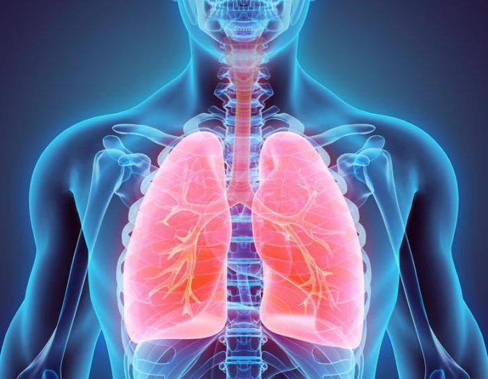
The thoracic cavity is the space in the body that is located inside. The thoracic and abdominal cavities separate the internal organs that they contain from the skeleton and muscles of the body, allowing these organs to move smoothly inside relative to the walls of the body. Organs located in the chest cavity: heart, blood vessels and nerves, trachea, bronchi and lungs; The esophagus passes from the chest cavity to the abdominal cavity through an opening in the diaphragm. The abdominal cavity contains the stomach and intestines, liver, kidneys, spleen, pancreas, numerous blood vessels and nerves.
The photo shows where and what organs of the chest cavity are located. Heart, trachea, esophagus, thymus, large vessels and the nerves are located in the space between the lungs - in the so-called mediastinum. Dome diaphragm, attached to the lower ribs, back of the sternum and lumbar vertebrae, forms a barrier between the human thoracic and abdominal organs.
Heart
The most worked muscle human body- heart or myocardium. The heart steadily, with a certain rhythm, without stopping, distils blood - approximately 7200 liters daily. Different parts of the myocardium synchronously contract and relax with a frequency of approximately 70 times per minute. During intense physical work, the load on the myocardium can triple. Heart contractions are started automatically - by a natural pacemaker located in its sinoatrial node.

The myocardium works automatically and is not subject to consciousness. It is formed by many short fibers - cardiomyocytes, interconnected into a single system. Its work is coordinated by a system of conductive muscle fibers consisting of two nodes, one of which contains the center of rhythmic self-excitation - the pacemaker. It sets the rhythm of contractions, which can change under the influence of nerve and hormonal signals from other parts of the body. For example, under heavy workload the heart beats faster, sending more blood to the muscles per unit of time. Thanks to its efficiency, about 250 million liters of blood are passed through the body over 70 years of life.
Trachea
This is the first of the organs of the human chest cavity. This organ is designed to pass air into the lungs and is located in front of the esophagus. The trachea originates at the height of the sixth cervical vertebra from the cartilage of the larynx and branches into bronchi at the height of the first thoracic vertebra.
The trachea is a tube 10-12 cm long and 2 cm wide, consisting of two dozen horseshoe-shaped cartilages. These cartilaginous rings are held in place anteriorly and laterally by ligaments. The space of each horseshoe ring is filled with connective tissue and smooth muscle fibers. The esophagus is located immediately behind the trachea. Inside, the surface of this organ is covered with a mucous membrane. The trachea, dividing, forms the following organs of the human chest cavity: the right and left main bronchi, descending into the roots of the lungs.

Bronchial tree
The branching in the shape of a tree contains the main bronchi - right and left, partial bronchi, zonal, segmental and subsegmental, small and terminal bronchioles, behind them are the respiratory sections of the lungs. The structure of the bronchi varies throughout the bronchial tree. The right bronchus is wider and placed more steeply downward than the left bronchus. Above the left main bronchus is the aortic arch, and below and in front of it is the aorta, which divides into two pulmonary arteries.
Structure of the bronchi
The main bronchi diverge to create 5 lobar bronchi. From them further go 10 segmental bronchi, each time decreasing in diameter. Smallest branches bronchial tree- bronchioles with a diameter of less than 1 mm. Unlike the trachea and bronchi, bronchioles do not contain cartilage tissue. They consist of many smooth muscle fibers, and their lumen remains open due to the tension of elastic fibers.
The main bronchi are located perpendicularly and rush towards the gates of the corresponding lungs. At the same time, the left bronchus is almost twice as long as the right one, has 3-4 more cartilaginous rings than the right bronchus, and seems to be a continuation of the trachea. The mucous membrane of these organs of the thoracic cavity is similar in structure to the mucous membrane of the trachea.

The bronchi are responsible for the passage of air from the trachea to the alveoli and back, as well as purifying the air from foreign impurities and removing them from the body. Large particles leave the bronchi during a cough. And small particles of dust or bacteria that have penetrated into respiratory organs chest cavity, are excreted by the movements of the cilia of epithelial cells, promoting bronchial secretion towards the trachea.
Lungs
In the chest cavity there are organs that everyone calls the lungs. This is the main one paired organ breathing, which occupies most of the chest space. The right and left lungs are divided according to location. In their shape, they resemble cut cones, with the apex directed towards the neck, and the concave base towards the diaphragm.
The top of the lung is 3-4 cm above the first rib. External surface adjacent to the ribs. The bronchi lead to the lungs, pulmonary artery, pulmonary veins, and nerves. The entry point of these organs is called the portal of the lung. Right lung has a greater width, but is shorter than the left one. The left lung has a niche in the lower front part for the heart. The lung contains a significant amount connective tissue. It has very high elasticity and helps the contractile forces of the lungs, which are necessary with every inhalation and exhalation.
Lung capacity
At rest, the volume of inhaled and exhaled air is on average about 0.5 liters. The vital capacity of the lungs, that is, the volume at the deepest exhalation after the most take a deep breath is in the range from 3.5 to 4.5 liters. For an adult, the rate of air consumption per minute is approximately 8 liters.
Diaphragm
The respiratory muscles rhythmically increase and decrease the volume of the lungs, changing the size of the chest cavity. The main work is done by the diaphragm. As it contracts, it flattens and descends, increasing the size of the chest cavity. The pressure in it drops, the lungs expand and draw in air. This is also facilitated by the raising of the ribs by the external intercostal muscles. Deep and rapid breathing involves accessory muscles, including the pectoral and abdominal muscles.

The mucous membrane of these organs of the chest cavity consists of epithelium, which, in turn, consists of many. The epithelium of the branches of the bronchial tree contains many endocrine cells that control the blood supply to the lungs and maintain the tone of the bronchial muscles.
To summarize all of the above, it is necessary to note that the organs of the human chest cavity are the basis of his life. It is impossible to live without a heart or lungs, and disruption of their functioning leads to serious illnesses. But human body- a perfect mechanism, you just need to listen to its signals and not harm, but help Mother Nature in its treatment and restoration.
The chest is part of the external respiratory apparatus. It performs supporting, motor, protective function.
Rib cage. Structure
This area is represented by a structure that has an osteochondral skeleton. Lymphatic and blood vessels, corresponding skeletal muscles, other soft fibers.
The osteochondral skeleton consists of twelve thoracic vertebrae, twelve pairs of ribs and the sternum. They are connected to each other through various types connections.
The cavity of the structure contains internal organs: lungs, lower respiratory tract, esophagus, heart and others.
The chest is presented in the shape of an irregular cone, the top of which is cut off. It defines four walls. The anterior one is formed by the costal cartilages and sternum, the posterior one by the posterior edges of the ribs and the thoracic vertebrae. The lateral (lateral) walls are formed by ribs, which are separated by intercostal spaces (intercostal spaces).
The thorax has an upper aperture (opening), limited by the first upper end with the jugular notch located on it and the inner ends of the first ribs. The hole is inclined anteriorly. The leading edge of the aperture is lowered downwards in the direction of the ribs. Thus, the jugular notch in the sternum is located between the second and third thoracic vertebrae at the level intervertebral disc.
The esophagus and trachea pass through the upper opening.
The inferior foramen is bounded by the body of the twelfth thoracic vertebra posteriorly, the sternal xiphoid process anteriorly, and the lower ribs on the sides. Its size is significantly larger than the size of the upper aperture.
The junction of the seventh to tenth costal pairs forms the anterolateral margin (costal arch). The left and right costal arches laterally limit the substernal angle, which is open downward. At its apex, located at the level of the ninth thoracic vertebra, there is
A diaphragm having an opening for the passage of the esophagus, aorta, inferior vein, closes the lower aperture.
Pulmonary grooves are located on the sides of the thoracic vertebrae. In them, the posterior portions of the lungs are adjacent to the walls of the chest.
Flexible rib arches give elasticity and greater strength to the entire structure.
The chest can have different shapes and sizes.
The movement of the entire structure is determined by the processes of exhalation and inhalation (breathing movements). Due to the fact that the anterior ends of the ribs are connected to the sternum, inhalation is accompanied by movement of both the sternum and ribs. Their elevation leads to an increase in the anteroposterior (sagittal) and transverse dimensions of the cell, and expansion of the intercostal spaces (intercostal spaces). All these factors explain the increase in cavity volume.
Exhalation is accompanied by drooping of the sternum and ends of the ribs, a significant decrease in the anteroposterior size, and narrowing of the intercostal spaces. All this leads to a decrease in cavity volume.
Chest deformity
This phenomenon often occurs in children. The two most common are funnel breast and chicken breast.
In the first case, the condition is caused by an abnormal inward retraction of the sternum. Chicken breast is when the chest sticks out. It should be noted that this type deformities are detected quite rarely in practice.
Structural anomalies certainly affect the child’s health. With a protruding chest, emphysema often develops ( chronic illness lungs, manifested by breathing problems).
As practice shows, in most cases this type of deformity requires surgical intervention.