Levator superioris muscle latin. Diseases of the eyelid muscles. Ideal eye proportions
Contents of the article: classList.toggle()">toggle
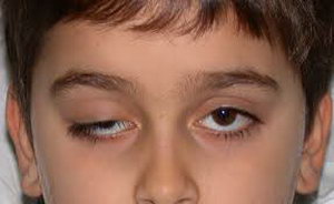
Ptosis of the eyelid is a pathology of location upper eyelid, in which it is lowered down and partially or completely covers the palpebral fissure. Another name for the anomaly is blepharoptosis.
Normally, the eyelid should overlap the iris of the eye by no more than 1.5 mm. If this value is exceeded, they speak of pathological drooping of the upper eyelid.
Ptosis is not only a cosmetic defect that significantly distorts appearance person. It interferes with the normal functioning of the visual analyzer, as it interferes with refraction.
Classification and causes of eyelid ptosis
Depending on the moment of occurrence, ptosis is divided into:
- Acquired
- Congenital.
Depending on the degree of drooping of the eyelid, it happens:
- Partial: covers no more than 1/3 of the pupil
- Incomplete: covers up to 1/2 of the pupil
- Full: The eyelid completely covers the pupil.
The acquired type of the disease, depending on the etiology (the cause of the appearance of ptosis of the upper eyelid), is divided into several types:

Regarding cases congenital ptosis, then it can arise due to two reasons:
- Anomaly in the development of the muscle that lifts the upper eyelid. May be combined with strabismus or amblyopia (lazy eye syndrome).
- Damage to the nerve centers of the oculomotor or facial nerve.
Symptoms of ptosis
The main clinical manifestation of the disease is drooping of the upper eyelid, which leads to partial or complete closure of the palpebral fissure. At the same time, people try to tense the frontalis muscle as much as possible so that the eyebrows rise and the eyelid stretches upward.
For this purpose, some patients throw back their heads and take a specific pose, which in the literature is called the stargazer pose.
A drooping eyelid prevents blinking movements, which leads to soreness and eye fatigue. A decrease in blink frequency causes tear film damage and development. Infection of the eye and development of an inflammatory disease can also occur.
Features of the disease in children
 Ptosis is difficult to diagnose in infancy. This is largely due to the fact that most of the time the child sleeps and is with eyes closed. You need to carefully monitor the baby's facial expression. Sometimes the disease may manifest as frequent blinking of the affected eye during feeding.
At an older age, ptosis in children can be suspected by the following signs:
- While reading or writing, the child tries to throw back his head. This is due to the limitation of visual fields when the upper eyelid droops.
- Uncontrolled muscle contraction on the affected side. Sometimes this is mistaken for a nervous tic.
- Complaints about rapid fatigue after visual work.
Cases of congenital ptosis may be accompanied by epicanthus(overhanging folds of skin over the eyelid), damage to the cornea and paralysis of the oculomotor muscles. If ptosis in a child is not eliminated, it will lead to development and decreased vision.
Diagnostics
A routine examination is sufficient to diagnose this disease. To determine its degree, it is necessary to calculate the MRD indicator - the distance between the center of the pupil and the edge of the upper eyelid. If the eyelid crosses the middle of the pupil, then the MRD is 0, if higher, then from +1 to +5, if lower, from -1 to -5.A comprehensive examination includes the following studies:
- Determination of visual acuity;
- Determination of visual fields;
- Ophthalmoscopy with examination of the fundus;
- Examination of the cornea;
- Study of tear fluid production;
- Biomicroscopy of the eyes with assessment of the tear film.
It is very important that while determining the extent of the disease, the patient is relaxed and does not frown. Otherwise, the result will be unreliable.
Children are examined especially carefully, since ptosis is often combined with eye amblyopia. Be sure to check visual acuity using Orlova's tables.
Treatment of ptosis
Elimination of ptosis of the upper eyelid can only be done after determining the root cause
Treatment of ptosis of the upper eyelid is possible only after determining the root cause. If it is neurogenic or traumatic in nature, its treatment necessarily includes physical therapy: UHF, galvanization, electrophoresis, paraffin therapy.
Operation
As for cases of congenital ptosis of the upper eyelid, it is necessary to resort to surgical intervention. It is aimed at shortening the muscle that lifts the eyelid.
Main stages of the operation:

The operation is also indicated if the upper eyelid still remains drooping after treatment of the underlying disease.
After the intervention, an aseptic (sterile) bandage is applied to the eye and prescribed antibacterial drugs wide range actions. This is necessary to prevent wound infection.
Medicine
Drooping upper eyelid can be treated with conservative methods. To restore the functionality of the extraocular muscles, the following therapy methods are used:

If the upper eyelid droops after a botulinum injection, then it is necessary to instill eye drops with alphagan, ipratropium, lopidine, and phenylephrine. Such drugs promote contraction of the extraocular muscles and, as a result, the eyelid rises.
You can speed up the lifting of the eyelid after Botox with the help of medical masks and creams for the skin around the eyelids. Professionals also recommend massaging your eyelids daily and visiting a steam sauna.
Exercises
A special gymnastic complex helps strengthen and tighten the extraocular muscles. This is especially true for involutional ptosis, which occurs as a result of natural aging.
Gymnastics for the eyes with ptosis of the upper eyelid:

Only with regular performance of a set of exercises for ptosis of the upper eyelid will you notice the effect.
Folk remedies
Treatment of ptosis of the upper eyelid, especially on initial stage, perhaps at home. Folk remedies are safe, and side effects practically absent.
Folk recipes for combating ptosis of the upper eyelid:

With regular use folk remedies not only strengthen muscle tissue, but also smooth out small wrinkles.
Amazing results can be achieved with the combined use of masks and massage. Massage technique:
- Treat your hands with an antibacterial agent;
- Remove makeup from the skin around the eyes;
- Treat your eyelids with massage oil;
- Perform light stroking movements on the upper eyelid in the direction from the inner corner of the eye to the outer. When treating the lower eyelid, move in the opposite direction;
- After warming up, lightly tap the skin around the eyes for 60 seconds;
- Then continuously press on the skin of the upper eyelid. Do not touch your eyeballs when doing this;
- Cover your eyes with cotton pads soaked in chamomile infusion.
Photo of ptosis of the upper eyelid


This also includes the muscle that lifts the upper eyelid (m. levator palpebrae superioris).
Start : thin narrow tendon fixed to the lesser wing sphenoid bone above the common tendon ring of Zinn and above and outside the optic foramen.
Attachment : orbital septum 2-3 mm above the edge of the cartilage (8-10 mm from the edge of the eyelid).
Blood supply : superior (lateral) muscular artery (branch ophthalmic artery), supraorbital artery, posterior ethmoidal artery, peripheral arterial arch of the upper eyelid.
Innervation : bilateral through the superior branch of the oculomotor nerve (n. III). Upper branch n. III enters the levator from below at the border of its posterior and middle thirds - 12–13 mm from the apex of the orbit.
Anatomy details : abdominal length - 40 mm, aponeurosis - 20–40 mm.
Three servings of muscle:
- The middle muscle portion, consisting here of a thin layer of smooth fibers (rostio media; m. tarsalis superior s. m. H. Mulleri), is woven into the upper edge of the cartilage; this portion is innervated by the cervical sympathetic nerve, while the remaining mass of striated levator fibers receives innervation from the oculomotor nerve.
- The anterior portion of the levator ending, turning into a wide aponeurosis, is directed to the tarso-orbital fascia; slightly below the superior orbital-palpebral groove it penetrates in separate bundles through this fascia, reaches the anterior surface of the cartilage and spreads all the way to the skin of the eyelid.
- Finally, the third, posterior, portion of the levator (also tendon) is directed to the upper fornix of the conjunctiva.
Such a triple ending of the muscle that lifts the upper eyelid, during its contraction, provides the possibility of joint movement of the upper eyelid as a whole through the cartilage (middle portion), the skin of the upper eyelid (anterior portion) and the upper conjunctival fornix (posterior portion of the muscle).
With normal levator tone, the upper eyelid occupies such a position that its edge covers the cornea by about 2 mm. Dysfunction of the elevator is expressed by the main symptom - drooping of the upper eyelid (ptosis) and, in addition, smoothness of the superior orbital-palpebral groove.
In the lower eyelid, there is no formalized muscle similar to the levator, i.e., the “descender” of the eyelid. However, the lower eyelid is pulled back when the eye turns downward by fascial processes that penetrate into the thickness of the eyelid and into the lower transitional fold of the conjunctiva from the sheath of the inferior rectus muscle eyeball. These cords, to which smooth muscle fibers may be mixed, are then given by some authors the name m. tarsalis inferior.
The course of the muscle is located lateral to the superior oblique and over the superior rectus muscle. In the anterior part of the upper part of the orbit, the levator is surrounded by a thin layer of fatty tissue, and here it is accompanied by the superior orbital artery, frontal and trochlear nerves, separating it from the roof of the orbit.
The superior rectus and levator of the upper eyelid are easily separated, despite their close proximity, except for their medial part, where they are connected by a fascial membrane. Both muscles originate from the same area of mesoderm. Both muscles are innervated by the superior branch of the oculomotor nerve. The nerve penetrates the muscles from the lower side at a distance of 12-13 mm from the apex of the orbit. Usually the nerve trunk approaches the levator from the outside of the superior rectus muscle, but it can also pierce it.
Directly behind the superior edge of the orbit, a section of dense tissue is attached to the levator superiorly. fibrous tissue(superior transverse ligament of Withnell, supporting the eyeball). The connection between them is quite strong, especially in the outer and inner parts. In this regard, their separation is possible only in the central areas. On the medial side, the Withnell ligament ends near the trochlea, while it passes in the form of fibrous cords under the superior oblique muscle of the eye behind, mixing with the fascia covering the supraorbital recess. From the outside, the ligament of Withnell connects to fibrous capsule lacrimal gland and periosteum of the frontal bone.
Withnell suggests that the main function of this ligament is to limit posterior displacement (tension) of the muscle. The author put forward this assumption due to the fact that its localization and distribution are similar to the limiting ligaments of the external muscles of the eye. The tension of the ligament provides support for the upper eyelid. If the ligament is destroyed, the levator of the upper eyelid sharply thickens and with inside ptosis occurs.
The distance from the transverse ligament of Withnell to the lower edge of the cartilaginous plate is 14-20 mm, and from the levator aponeurosis to the circular and skin insert is 7 mm.
In addition to the palpebral insertion, the levator aponeurosis forms a wide fibrous cord that attaches to the edge of the orbit immediately behind the internal and external eyelid ligaments. They are called the inner "horn" and the outer "horn". Since they are quite rigid, during levator resection it is possible to maintain the upper eyelid in the desired position by fixing the “horn” with an instrument.
The outer “horn” is a rather powerful bundle of fibrous tissue that partially divides the inner part of the lacrimal gland into two parts. It is located below, attaching in the area of the outer tubercle of the orbit to the outer ligament of the eyelid. Not taking this into account anatomical feature during removal of a tumor of the lacrimal gland can lead to ptosis of the lateral part of the upper eyelid. The internal "horn", on the contrary, becomes thinner, turns into a thin film that passes over the tendon of the superior oblique muscle towards the internal ligament of the eyelid and the posterior lacrimal crest.
The fibers of the levator tendon are woven into the connective tissue of the cartilaginous plate of the upper eyelid approximately at the level of its upper third. When the muscle contracts, the eyelid rises and at the same time the preaponeurotic space is shortened and the postaponeurotic space is lengthened.
4644 0
The eyelids are movable structures that protect the eyeball from the front. There are upper (palpebra superior) and lower (palpebra inferior) eyelids. Thanks to the mobility of the eyelids, namely due to their blinking, the tear fluid is evenly distributed over the front surface of the eye, moisturizing the cornea and conjunctiva. The connection of the upper and lower eyelids occurs through the medial commissure (commissura medialis palpebrarum) and the lateral commissure (commissura lateralis palpebrarum), which begin respectively in the outer (angulus oculi lateralis) and inner corner of the eye (angulus oculi medialis).
In the inner corner, at a distance of approximately 5 mm before the junction of the eyelids, a recess is formed - the lacrimal lake (lacus lacrimalis). At its bottom there is a rounded pink tubercle - the lacrimal caruncle (caruncula lacrimalis), to which is adjacent the semilunar fold of the conjunctiva (plica semilunaris conjunctivae). The almond-shaped space between the open eyelids is called the palpebral fissure (rima palpebrarum). Its horizontal length in an adult is 30 mm, and its height in the center is from 10 to 14 mm. When the eyelids are closed, the palpebral fissure completely disappears.
In the eyelids, two plates are conventionally distinguished - the outer (musculocutaneous) and the inner (conjunctival-cartilaginous). The skin of the eyelids contains sebaceous sweat glands. The subcutaneous tissue of the eyelids is devoid of fat, so swelling and hemorrhages easily spread in it, it easily folds, forming upper and lower folds that coincide with the corresponding edges of the cartilage. The cartilages of the eyelids (tarsus superior et inferior) look like a slightly convex plate about 20 mm long, up to 12 mm high and about 1 mm thick. The height of the cartilage on the lower eyelid is 5-6 mm; on the upper eyelid the cartilage is more pronounced. Cartilage consists of dense connective tissue and does not have its own cartilage cells. They are connected to the upper and lower walls of the orbit by ligaments of the eyelids (lig. palpebrale mediale et laterale).
The orbital part of the cartilage is connected to the edges of the orbit through dense fascia (septum orbitale). The cartilages contain elongated alveolar glands (glandulae tarsales), about 20 of them in the lower eyelid and 25 in the upper. The glands are located in parallel rows, their excretory ducts open near the posterior free edge of the eyelids. The lipid secretion of the glands lubricates the intercostal space of the eyelids, forming the outer layer of the precorneal tear film, which prevents tears from rolling down through the lower edge of the eyelid.
The connective tissue membrane (conjunctiva) covering the back surface of the eyelids is tightly fused with cartilage. When the conjunctiva passes from the eyelids to the eyeball, it forms movable vaults - upper and lower. The edges of the eyelids, forming the palpebral fissure, are limited in front by the anterior rib, and behind by the posterior rib. The narrow strip between them, up to 2 mm wide, is called the intercostal (intermarginal) space; here the roots of the eyelashes are located in 2-3 rows, sebaceous glands(Glands of Zeiss), modified sweat glands (Glands of Moll), openings of the excretory ducts of the Meibomian glands. At the inner corner of the eye, the intermarginal space narrows and passes into the lacrimal papilla (papilla lacrimalis), at the top of which there is an opening - punctum(punctum lacrimale); it is immersed in the lacrimal lake and opens into the lacrimal canaliculus (canaliculus lacimalis).
Eyelid muscles
Under the skin of the eyelids, ensuring their mobility, there are two groups of muscles - antagonists in the direction of action: the circular muscle of the eye (m. orbicularis oculi) and the muscle that lifts the upper eyelid (m. levator palpebrae superioris).
Orbicularis oculi muscle consists of the following parts: orbital (pars orbitalis), palpebral, or age-old (pars palpebralis), and lacrimal (pars lacrimalis). The orbital part is a circular belt, the fibers of which are attached to the medial ligament of the eyelids (lig. parpebrale mediale) and the frontal process of the maxilla. When this part contracts, the eyelids close tightly. The fibers of the palpebral part begin from the medial ligament of the eyelids and, forming an arc, reach the outer corner of the eye, attaching to the lateral ligament of the eyelids. When this muscle group contracts, the eyelids close and blink.
The lacrimal part is a group of muscle fibers that start from the posterior lacrimal crest of the lacrimal bone (os lacrimalis), then pass behind the lacrimal sac (saccus lacrimalis), intertwining with the fibers of the palpebral part. The muscle fibers enclose the lacrimal sac in a loop, as a result of which, when the muscle contracts, the lumen of the lacrimal sac either expands or narrows. Thanks to this, the process of absorption and movement of tear fluid along the lacrimal ducts occurs.
There are muscle fibers of the orbicularis oculi muscle, which are located between the roots of the eyelashes around the duct of the meibomian glands (m. ciliaris Riolani). Contraction of the fibers promotes the secretion of the mentioned glands and a tight fit of the edge of the eyelids to the eyeball. The circular muscle is innervated by the zygomatic (rr. zygomatici) and temporal (rr. temporales) branches of the facial nerve.
Levator superioris muscle, begins near the optic canal (canalis opticus), goes under top part orbit and ends in three muscle plates. The superficial plate, forming a wide aponeurosis, perforates the tarso-orbital fascia and ends above the skin of the eyelid. The middle one consists of a thin layer of smooth fibers (m. tarsalis superior, m. Mulleri), intertwined with the upper edge of the cartilage, innervated by sympathetic nerve fibers. A deep plate in the form of a wide tendon reaches the upper fornix of the conjunctiva and is attached there. The superficial and deep plates are innervated by the oculomotor nerve.
The lower eyelid is retracted muscle of the lower eyelid cartilage(m. tarsalis inferior) and fascial processes of the inferior rectus muscle (m. rectus inferior).
Blood supply
The blood supply to the eyelids is carried out through the branches of the ophthalmic artery (a. ophthalmica), which is part of the internal system carotid artery, as well as anastomoses from the facial and maxillary arteries (aa. facialis et maxiaJlaris) from the external carotid artery system. These arteries branch and form arterial arches: two on the upper eyelid, one on the lower. The arteries correspond to veins, through which the outflow of venous blood occurs mainly towards the angular vein (v. angularis), vein of the lacrimal gland (v. lacrnnalis) and temporal superficial vein(v. temporalis superfirialis). The structural features of these veins include the absence of valves and the presence large quantity anastomoses. It is clear that such features can cause the development of severe intracranial complications, for example, with the development of purulent processes on the face.
Lymphatic system
The lymphatic network is well developed on the eyelids; There are two levels, which are located on the anterior and posterior surfaces of the cartilage. Lymphatic vessels upper eyelids flow into preauricular The lymph nodes, lower eyelid - into the submandibular lymph nodes.
Innervation
Branches of the facial nerve (n. facialis) and three branches trigeminal nerve(n. trigeminus), as well as the large auricular nerve (n. auricularis majos) provide sensitive innervation to the skin of the face. The skin and conjunctiva of the eyelid are innervated by two main branches of the maxillary nerve (n. maxillaris) - the infraorbital (n. infraorbitalis) and zygomatic (n. zygomaticus) nerve.
Eyelid research methods
To study the condition of the eyelids, the following research methods are used:
1. External examination of the eyelids, palpation.
2. Inspection with side (focal) lighting.
3. Inspection of the mucous membrane of the eyelids when everting the upper and lower eyelids.
4. Biomicroscopy.
Diseases of the eyelids
Among the total number of patients with inflammatory diseases 23.3% of eyes are patients with inflammation of the eyelids. Pathology of the auxiliary and protective apparatus of the eyes is of great socio-economic importance, as it is one of the most common causes of temporary disability and can lead to significant complications in the visual organ.
Zhaboyedov G.D., Skripnik R.L., Baran T.V.
Ptosis (drooping) of the upper eyelid is an uncontrolled disruption of the muscles that raise and lower the upper eyelid. Muscle weakness is expressed as a cosmetic defect in the form of asymmetry in the size of the palpebral fissures, which develops into a host of complications, including loss of vision.
The disease affects patients of any age, from newborns to pensioners. All treatment methods, including the main surgical therapy for ptosis, are aimed at increasing the tone of the eye muscles.

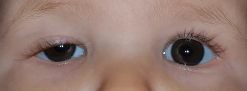
Blepharoptosis (drooping of the upper eyelid) is a pathology of the muscular system in which the eyelid partially or completely covers the iris or pupil, and in advanced stages, completely covers the palpebral fissure. Normally, the right and left eyelids should cover no more than 1.5-2 mm of the upper edge of the iris. If the muscles are weak, poorly innervated, or damaged, the eyelid loses control and droops below normal.
Ptosis is a disease of only the upper eyelid, since the lower eyelid lacks the levator muscle, which is responsible for lifting. There is a small Müller muscle located there, which is innervated in cervical spine and is only capable of widening the palpebral fissure by a couple of millimeters. Therefore, with paralysis sympathetic nerve, which is responsible for this small muscle on the lower eyelid, ptosis will be slight, completely unnoticeable.

Physical obstruction of the visual field leads to a number of complications that are especially dangerous in childhood when the visual function is just developing. Ptosis in a child leads to developmental disorders binocular vision, .
All these complications are typical for adults, but when they appear in infant contribute to the incorrect training of the brain to compare visual images. Subsequently, this will lead to the impossibility of correcting or restoring correct vision.
Classification and reasons
Muscle weakness can be acquired or congenital. Congenital ptosis of the upper eyelid is a disease of young children, its causes are underdevelopment or absence of the muscles that lift the eyelid, as well as damage to the nerve centers. Congenital ptosis is characterized by bilateral damage to the upper eyelid of the right and left eyes simultaneously.
Look interesting video O congenital form diseases and methods of treatment:
Unilateral lesions are characteristic of acquired ptosis. This type of ptosis develops as a complication of another, more serious pathological process.
Classification of ptosis of the upper eyelid depending on the cause of its appearance:
- Aponeurotic blepharoptosis – excessive stretching or relaxation of muscles, loss of tone.
- Neurogenic ptosis is a violation of the passage of nerve impulses to control muscles. Neurogenic ptosis is a symptom of a central nervous system disease; the appearance of neurology is the first signal for additional examination of brain structures.
- Mechanical blepharoptosis is post-traumatic muscle damage, tumor growth, and scarring.
- Age - natural physiological processes aging of the body provokes weakening and stretching of muscles and ligaments.
- False blepharoptosis – observed with a large volume of skin folds.
Other causes of blepharoptosis in adults include:
- damage, bruises, ruptures, eye injuries;
- diseases nervous system or brain: stroke, neuritis, multiple sclerosis, tumors, neoplasms, hemorrhages, aneurysms, encephalopathy, meningitis, cerebral palsy;
- paresis, paralysis, ruptures, muscle weakness;
- diabetes mellitus or other endocrine diseases;
- exophthalmos;
- a consequence of unsuccessful plastic surgery, Botox injections.
By stages:
- partial;
- incomplete;
- full.

Ptosis has 3 degrees, which are measured in the number of millimeters of distance between the edge of the eyelid and the center of the pupil. In this case, the patient’s eyes and eyebrows should be relaxed and in a natural position. If the location of the edge of the upper eyelid coincides with the center of the pupil, this is the equator, 0 millimeters.
Degrees of ptosis:
- First degree – from +2 to +5 mm.
- Second degree – from +2 to -2 mm.
- Third degree – from –2 to –5 mm.
Symptoms of the disease
Eyelid ptosis is characterized by the main, most obvious visual symptom - drooping with a partially or completely closed palpebral fissure. At the early stage of the disease, pay attention to the symmetry of the location of the eyelids of the right and left eyes relative to the edge of the cornea.
Other manifestations of blepharoptosis:
- decreased visual acuity in one eye;
- fast fatiguability;
- astrologer pose, when the patient has to throw his head back to get a clear image;
- double vision;
- the pathological eye stops blinking, this leads to;
- the resulting pocket under the drooping eyelid contributes to the accumulation of bacteria, subsequently the development of frequent inflammation;
- double vision;
- unconsciously the patient tries to lift the upper eyelid with brow ridges or forehead muscles;
- gradual development of strabismus.

Diagnostics
Diagnostics is aimed at identifying the root cause of the disease and prescribing adequate treatment. drooping eyelid early stages hardly noticeable, but it's extremely important sign beginning of development serious illnesses, such as a brain tumor. Therefore, it is important for the ophthalmologist to find out whether ptosis is congenital or appears suddenly. To do this, the patient is interviewed and an anamnesis is collected.
It happens that the patient has not noticed the prolapse before or cannot say exactly when it appeared. In this case, it is necessary to conduct additional examinations to exclude all possible causes of the disease.
Stages of diagnosing blepharoptosis:
- Visual inspection, measurement of the degree of ptosis.
- Measurement of acuity, visual field, intraocular pressure, fundus examination.
- Biomicroscopy of the eye.
- Measurement of muscle tone, fold symmetry and blinking.
- Ultrasound of the eye, electromyography.
- Radiography.
- MRI of the head.
- Checking for binocular vision.
- Examination by a neurosurgeon, neurologist, endocrinologist.
How to cure upper eyelid ptosis
It is necessary to fight ptosis only after finding out the cause. In the early stages congenital pathology in the absence of visual impairment or a small cosmetic defect, it is recommended not to treat, but to carry out comprehensive prevention.
Treatment of ptosis is divided into conservative and surgical. Conservative methods goes well with homemade folk recipes.
For ptosis due to injury or nerve dysfunction, it is recommended to wait about a year after the incident. During this time, effective treatment can restore all nerve connections without surgery or significantly reduce its volume.

What to do if your eyelid droops after Botox
Botox (botulinum toxin) is medicine, derived from botulinum bacteria, which disrupts the neuromuscular connection. The drug contains a neurotoxin, which in small dosages, when applied locally, attacks and kills nerve cells in the muscles, due to which they completely relax.
When using the drug in the cosmetic industry, a complication of incorrect or inaccurate administration can be ptosis of the upper eyelid after Botox injection, the treatment of which is very long. Moreover, the first few procedures can be successful, but each subsequent one requires an increase in the amount of the drug, which can lead to an overdose, as the body learns to develop immunity and antibodies to botulinum toxin.
Removing prolapse (blepharoptosis) is difficult, but possible. The first option for the simplest non-surgical treatment is to do nothing or just wait. After about 2-3 months, the body will build additional lateral branches of the nerves, which will allow it to regain control of the muscle on its own.
The second method helps speed up this process; for this purpose, physiotherapeutic procedures are actively used (UHF, electrophoresis, massage, darsonval, microcurrents, galvanotherapy), proserin injections, large doses B vitamins, neuroprotectors. All this accelerates the restoration of innervation and promotes rapid resorption of Botox residues.
Operation
Surgery to correct ptosis (drooping) of the upper eyelid is called blepharoplasty. The operation is indicated in cases of advanced ptosis with impaired quality of vision. The intervention is performed under local anesthesia on an outpatient basis. The rehabilitation period lasts about a month, during which the patient is observed by the operating surgeon.

There are many methods of operation, but the essence is the same - to shorten the relaxed muscle either by cutting and removing a part, or by folding it in half and stitching it. Cosmetic seam hides in natural skin fold, and over time it completely resolves.
The cost of the operation depends on:
- complexity of the operation;
- stages of ptosis;
- additional research;
- the medical institution you have chosen;
- number of specialist consultations;
- number of laboratory diagnostics;
- type of anesthesia;
- accompanying pathologies.
On average, the amount per operation varies from 20 to 60 thousand rubles. You can find out the exact figure directly at your appointment, after examination by a specialist.
Watch the video to see how the operation (blepharoplasty) goes:
Home treatment
Ptosis of the upper eyelid can be treated conservatively at home. Treatment without surgery uses medications, massage, alternative medicine, physiotherapeutic procedures.
Methods for treating drooping eyelids using folk remedies:
- raw mask chicken egg with sesame oil, apply to the skin once a day, wash off with warm water;
- lotions or warm compresses from infusions of chamomile, calendula, rose hips, black tea, birch leaves;
- applying “dry heat” using a cloth bag with super-fried sea salt;
- grated potato mask raw potatoes applied for 20 minutes once a day;
- a mask of honey with aloe pulp is applied 2 times a day.
Traditional medications used internally, mainly B vitamins, neuroprotectors, drugs that stimulate growth, as well as regeneration of nerve tissue, enhancing nutrition nerve cells. Everything is prescribed individually and depends on the stage, form, and cause of ptosis.
Physiotherapy:
- vacuum massage for ptosis of the upper eyelid;
- electrophoresis;
- warming up;
- myostimulation with currents.
All procedures and medications must be clarified and agreed upon with your attending ophthalmologist. The information on the site is for informational purposes only; do not use it as a guide to action.
Additionally, we invite you to watch a video about ptosis. Elena Malysheva will tell you in detail about the disease and ways to combat it.
The key to good result when performing facial gymnastics and massages - precise knowledge of facial anatomy.
The fight against aging for a woman usually begins with the skin around the eyes, since this is where the first age-related problems appear: the skin loses its freshness, swelling and fine wrinkles appear.
And no wonder: in the eye area the layer of epidermis is very thin - only half a millimeter. In addition, there is almost no sebaceous glands, a “soft pad” of subcutaneous fat and very little muscle to maintain its elasticity. Collagen fibers (the “reinforcement” of the skin) are arranged here in the form of a mesh, so the skin of the eyelids is easily stretchable. And due to the looseness of the subcutaneous tissue, it is also prone to swelling. In addition, she is constantly in motion: her eyes blink, squint, and “smile.” As a result, the skin around the eyes is particularly stressed.
Therefore, let's start understanding the structure of the face from this area.
Anatomy of the area around the eyes
The eyelids and periorbital region are a single complex consisting of many anatomical structures that undergo changes during surgical manipulation
The skin of the eyelids is the thinnest on the body. The thickness of the eyelid skin is less than a millimeter.
Unlike other anatomical areas where fatty tissue lies under the skin, just under the skin of the eyelids lies the flat orbicularis oculi muscle, which is conventionally divided into three parts: internal, median and external.
The internal part of the orbicularis oculi muscle is located above the cartilaginous plates of the upper and lower eyelids, the middle part is above the intraorbital fat, the external part is located above the bones of the orbit and is woven above into the muscles of the forehead, and below into the superficial musculofascial system of the face (SMAS).
The orbicularis oculi muscle protects the eyeball, performs blinking, and functions as a “tear pump.”
The musculoskeletal system of the eyelids performs a supporting function and is represented by thin strips of cartilage - tarsal plates, lateral canthal tendons and numerous additional ligaments.
The superior tarsal plate is located on the lower edge of the upper eyelid under orbicularis muscle eyes, and is usually 30 mm long and 10 mm wide, it is firmly connected to internal part the orbicularis oculi muscle, the aponeurosis of the levator palpebrae superioris muscle, the Müller muscle and the conjunctiva. The inferior tarsal plate is located on top edge the lower eyelid, usually 28 mm long and 4 mm wide, is attached to the orbicularis muscle, capsulopalpebral fascia and conjunctiva. The lateral canthal tendons are located under the orbicularis oculi muscle and are firmly connected to it. They connect the tarsal plates to bony edges orbits.
Under the orbicularis muscle also lies the orbital septum - a thin but very strong membrane; one edge is woven into the periosteum of the bones surrounding the eyeball, and the other edge is woven into the skin of the eyelids. The orbital septum retains the intraorbital fat within the orbit.
Under the orbital septum there is intraorbital fat, which acts as a shock absorber and surrounds the eyeball on all sides.
Portions of the upper and lower intraorbital fat are divided into internal, central and external. Next to the upper outer portion is the lacrimal gland.
The muscle that lifts the upper eyelid opens the eye and is located in the upper eyelid under the cushion of fat. This muscle is attached to the superior tarsal cartilage.
The skin of the upper eyelid is usually attached to the levator palpebrae superioris muscle. At the site of attachment of the skin to this muscle when open eye a fold forms on the upper eyelid.
This supraorbital fold different people very different. In people from Asia, for example, it is weakly expressed or not at all; in Europeans, it is well expressed.
 |
|
| 1 - Müller muscle, 2 - Levator muscle of the upper eyelid 3 - Superior rectus muscle 4 - Inferior rectus muscle 5 - Inferior oblique muscle 6 - Orbital bones 7 - Edge of the eye socket 8 - SOOF - infraorbital fat 9 - Orbital ligament 10 - Orbital septum 11 - Intraorbital fat 12 - Capsulopalpebral fascia |
13 - Inferior pretarsal muscle 14 - Lower tarsal plate 15 - Superior pretarsal muscle 16 - Upper tarsal plate 17 - Conjunctiva 18 - Links 19 - Muscle that lifts the upper eyelid 20 - Orbital septum 21 - Intraorbital fat 22 - Eyebrow 23 - Eyebrow fat 24 - Bones of the orbit |
Behind these structures is the eyeball itself, which is supplied with blood and innervated through back eye sockets.
The muscles that move the eye are attached at one end to the eyeball and lie on its surface, and at the other end they are attached to the bones of the orbit.
The nerves that control the muscles are small branches of the facial nerve and enter the orbicularis oculi muscle on all sides from its outer edges.
The anatomical structures of the lower eyelid and midface are closely related, and changes in the anatomy of the midface affect the appearance of the lower eyelid. In addition to portions of periorbital fat, two additional layers of fatty tissue exist in the midface.

Beneath the outer part of the orbicularis oculi muscle lies the infraorbital fat (SOOF). The greatest thickness of SOOF is on the outside and sides.
The SOOF is deep to the superficial musculoaponeurotic system of the face (SMAS) and envelops the zygomatic major and minor muscles.
In addition to SOOF, malar fat pad is an accumulation of fat in the form of a triangle or so-called. "painting" fat is located under the skin, above the SMAS.
Aging of the midface is often accompanied by sagging of the malar fatty tissue, which results in noticeable zygomatic or so-called “painting” bags on the face.
The main supporting structure of the midface is the orbitozygomatic ligament, which runs from the bones almost along the edge of the orbit to the skin. It contributes to the formation of the zygomatic “painting” bag and the eyelid-cheek separation visible with age.
Ideal eye proportions
As a rule, a good aesthetic result is obtained only when the proportions of the eye and eyelids are in accordance with the proportions of the face. Outside, the eyelids and paraorbital region are represented by many anatomical structures.
The palpebral fissure is formed by the edge of the upper and lower eyelids. If you measure the eye, it usually measures 30-31 mm horizontally and 8-10 mm vertically.
The outer canthus is usually located 2 mm above the inner canthus in men and 4 mm in women, forming an inclination angle of 10-15 degrees, i.e. the palpebral fissure is slightly inclined from outside to inside and from top to bottom.
However, the position of the outer corner of the eye may change due to age and may be influenced by heredity, race, and gender.

The edge of the upper eyelid usually covers the iris by approximately 1.5 mm, and the lower eyelid begins immediately below the lower edge of the iris.
The normal position (protrusion) of the eyeball relative to bone walls orbits are observed in 65% of the population, and it ranges from 15 to 17 mm.
Deep-set eyes have a projection of less than 15 mm, and protruding eyes have a projection of more than 18 mm.

The size of the iris is approximately the same in all people, but the shape of the scleral triangles (triangles) white between the iris and the corners of the eye) may vary.
Typically, the nasal scleral triangle is smaller than the lateral one and has a more obtuse angle.
With increasing eyelid laxity and age, these triangles lose shape, especially the lateral scleral triangle.

The horizontal fold in the upper eyelid is formed by the aponeurosis of the levator palpebrae superioris muscle, which is woven into the skin, passing through the orbicularis oculi muscle.
Excess skin and muscle hangs over the crease, which is a fixed line. Both upper eyelid folds and the amount of skin overhanging them vary between individuals different races, they are influenced by gender and age.

The fold of the upper eyelid in Europeans is approximately 7 mm above the edge of the eyelid along a line drawn through the center of the pupil in men and 10 mm above the edge of the eyelid in women. IN lower eyelids ah, there are similar folds that are 2-3 mm below the edge of the eyelids. Typically, lower eyelid folds are more noticeable at a young age and less noticeable as you age. In Asians, the fold of the upper eyelid is either lower - no more than 3-4 mm above the edge of the eyelid or is absent.

Differences between the female and male eyes also appear in several other points: the inclination of the palpebral fissure (from the outside in and from top to bottom) in men is less pronounced than in women, the bone structures above the eye are more full and the eyebrow itself is usually wider, located lower and less curved.
Age-related changes in the upper and lower eyelids
The main features of young eyelids are a smooth contour extending from the eyebrow to the upper eyelid and from the lower eyelid to the cheek and midface. The eyelid-cheek division is located at the edge of the orbit and is usually 5-12 mm below the edge of the lower eyelid, the skin is taut and the tissues are full. From the inner canthus to the outer canthus, the horizontal axis of the eye has an upward slope.

In contrast, with age, the eyes appear hollow, with a clear boundary between the eyebrow and the upper eyelid, the lower eyelid and the cheek. In most people, the palpebral fissure becomes smaller and/or rounded with age due to the downward displacement of both the upper and lower eyelids. The eyelid-cheek division is located significantly below the edge of the orbit, 15-18 mm from the edge of the lower eyelid, and the slope from the inner canthus to the outer canthus becomes downward. Which gives the eyes a sadder look.
A youthful upper eyelid usually has minimal excess skin. Dermatochalasis, or excess skin, is a cardinal feature of the aging upper eyelid.

Constant contraction of the muscles surrounding the eyes, the creep of sagging forehead tissues and loss of elastic properties of the skin lead to the formation of the so-called. " crow's feet" - fan-shaped wrinkles located at the outer corner of the eye and fine wrinkles under the lower eyelid.
The youthful lower eyelid has a smooth, continuous transition zone between the eyelid and cheek without bulging orbital fat, indentation, or pigmentation.
With age, progressive skeletonization of the orbit occurs (the relief of the bones around the eye becomes more visible), since subcutaneous fat, covering the orbital frame atrophies and migrates downward. This downward displacement of fat results in loss of cheek convexity.
Also, pigmentation (darkening of the skin) or the so-called may appear on the lower eyelid. "circles under the eyes" with or without infraorbital depressions.
Eyelid bags or hernias can be caused by orbital weakening of the orbital septum, which stretches and causes orbital fat to protrude.

♦ Increase in length (height) of the lower eyelid
The nasolacrimal groove and zygomatic groove, which appear with age, can give the eye area an unaesthetic appearance. Atrophy of intraorbital fat associated with aging can make the eyes appear sunken and skeletal.
Many wrinkles around the eye may reflect loss of skin elasticity.

Aging of the eyelids. Causes and manifestations
Main reasons age-related changes in the eyelid area are stretching and weakening of the ligaments, muscles and skin of the face under the influence of gravitational forces - attraction. The elasticity of the facial ligaments weakens, they lengthen, but remain firmly fixed to the bones and skin.
Consequently, in the most mobile areas with minimal fixation of the ligaments to the skin, gravity pulls the tissue downward with the formation of protrusions. They are filled with deep ones adipose tissue, such as, " fatty hernias"lower or upper eyelid.
Where the ligaments hold the skin and muscles more firmly, depressions or grooves appear - relief folds.
In the area of the upper eyelids, these changes may look like overhang of skin and fatty tissue in the area of the outer corners of the eye (outer “bags” - Fig. 1) and inner corners of the eye (inner “bags” - Fig. 2), overhang of only the skin over the entire eyelid gap or only from the outside (dermatochalasis - Fig. 3), drooping of the entire upper eyelid (ptosis - Fig. 4).


In the area of the lower eyelids, these changes may look like drooping of the lower eyelid (exposure of the sclera - Fig. 5), an increase in the lower portion of the muscle surrounding the eyes (hypertrophy of the orbicularis oculi - Fig. 6), the appearance of “bags” under the eyes when intraorbital fat is no longer retained inside the orbit by the orbicularis oculi muscle and the orbital septum, losing their tone (“fatty hernias” - Fig. 7, Fig. 8).


♦ Classification of age-related changes in the eyelids
Age-related changes in the lower eyelid area develop over time and can be classified into the following four types:
 |
Type I- Changes are limited to the area of the lower eyelids; weakening of the muscle tone surrounding the eye and bulging of orbital fat may be observed. |
 |
Type II- Changes extend beyond the boundaries of the lower eyelids; weakening of the tone of the muscles surrounding the eyes, weakening of skin tone and the appearance of excess skin, slight drooping of the cheek tissue and the appearance of eyelid-cheek separation may be observed. |
 |
III type- Changes affect all tissues bordering the eyelids, lowering of the tissues of the cheeks and zygomatic region, increasing the separation of the eyelid-cheek, skeletonization of the orbit - the bones of the orbit become visible, the nasolabial folds deepen. |
 |
IV type- Further lowering of the eyelid-cheek separation, deepening of the nasolacrimal grooves, the appearance of the so-called. "malar" or zygomatic "bags", drooping of the outer corners of the eye and exposure of the sclera. |
This classification helps solve problems characteristic of each type of age-related changes in the eyelid area.
The classification demonstrates that the aging of the lower eyelid area and the midface area is inherently related to each other, and rejuvenation of one area without the other, in some cases, can lead to insufficient or unsatisfactory results.
It is important to note that one of the cornerstones of these changes is the real and obvious loss of tissue volume in the eyelids and cheeks, and only its restoration can sometimes improve the situation.