What blood test shows heart problems. My heart hurts, what tests should I take? Ultrasound of blood vessels, including lower extremities
Biochemical analysis is one of the most accessible, fastest and inexpensive examination methods. It allows you to assess the condition of the whole organism. Any disturbances in the functioning of internal organs invariably affect the composition of the blood. This analysis is used in all areas of medicine, including cardiology. Almost any medical examination begin with .
A biochemical blood test includes many indicators. Most often, only a part of these indicators is assigned, since a detailed analysis is required extremely rarely.
The essence of the analysis is to determine the amount and concentration chemical substances in blood. As you know, blood circulates in all tissues, therefore, when there is inflammation or disturbance in any of them, the composition of the blood also changes.
A biochemical therapeutic blood test can be prescribed if absolutely any disease is suspected. The examination is prescribed by therapists, hepatologists, gastroenterologists, and cardiologists.
- Confirmation of diagnosis. The analysis allows us to identify specific violations and clarify an existing diagnosis.
- Diseases of the liver, kidneys, and gastrointestinal tract. A biochemical therapeutic blood test is indispensable when examining the functioning of the gastrointestinal tract, since the indicators contain enzymes.
- Pregnancy. During pregnancy, blood tests are taken constantly, once every 2 weeks. The examination allows you to identify dysfunctions of internal organs, prevent miscarriage, and diagnose preeclampsia in a timely manner.
- Prevention. As preventive examination It is recommended to take a blood test annually. This will help detect early stage a whole range of diseases.
- Checking the effectiveness of treatment. If a diagnosis is made and treatment is prescribed, a blood test is prescribed during the treatment process or after completion of the course to check the progress and effectiveness of the prescribed treatment methods.
More information about the lipid profile can be found in the video:
Among the advantages of laboratory research are accessibility, low price, speed of the procedure, painlessness and high information content. The disadvantages include the possibility of error.
Blood reacts not only to internal changes, but also to external influences. Therefore, without proper preparation the result may be erroneous. Also, despite the high information content of the analysis, it is difficult to make an accurate diagnosis based on the results without further examination.
Cardiological profile: what tests are included in it

Cardiovascular diseases require particularly careful examination. On this moment this is the most common reason mortality among the population. For timely detection of diseases, an examination is prescribed.
A cardiac profile is a whole range of tests to check the condition of the heart and blood vessels. It is prescribed for any suspected malfunction cordially- vascular system and is initial stage examinations.
A cardiological profile allows not only to detect existing diseases, but also to determine the risk and likelihood of their occurrence, predict the course of the disease, and select treatment or preventive measures.With the help of a cardiological profile, it is possible to detect diseases at an early stage, in a latent form, when there are still no symptoms.
The cardiac profile includes the following tests:
- Lipidogram. This analysis allows you to determine the level of lipids in the blood, the tendency to. The indicators reveal disturbances in lipid metabolism. This includes cholesterol, HDL, LDL, triglycerides.
- . The analysis includes indicators. Coagulation disorders can lead to thrombosis or bleeding. Monitoring these indicators is necessary.
- AST. This enzyme is responsible for metabolism not only in liver tissue, but also in the heart muscle. The indicator is often used to diagnose myocardial infarction.
- Creatine kinase. This is an enzyme responsible for energy exchange in cells and tissues. If the level of this enzyme increases significantly, this indicates a risk of myocardial infarction.
- LDH. This enzyme is found in the heart muscle, kidneys, and liver tissues. Its level in the blood increases during myocardial infarction in the acute stage.
Indications for a cardiological profile are any heart disease, suspicion of myocardial infarction, and pain in chest, increased blood pressure.
Indicators of biochemical analysis and their application in cardiology

A complete biochemical analysis includes more than 20 indicators. Most often, the doctor specifies which indicators are necessary to make a diagnosis. The choice depends on the symptoms and the suspected disease.
When examining cardiovascular diseases, cardiac profile indicators are most often assessed. But other indicators may also be important in assessing the impact of heart disease.
The list of frequently prescribed biochemical blood test indicators includes:
- Glucose. People with suspected diabetes mellitus need to donate blood for sugar testing in order to control the metabolism in the body. Disorders of carbohydrate metabolism, as a rule, indicate a failure in endocrine system, as well as on various diseases liver.
- . Cholesterol has several varieties (high and low density lipids). Not all types of cholesterol are harmful to health. Everyone needs to monitor their cholesterol levels, especially people over 50 years of age, as the risk of atherosclerosis increases.
- Bilirubin. Bilirubin is a protein that is broken down in liver tissue. At large cluster bilirubin in the blood becomes toxic. This indicator is used to check the functioning of the liver and bile ducts.
- AST. An enzyme that applies to both liver tests and cardiological profile. Used in the diagnosis of heart attacks and liver diseases (cirrhosis, hepatitis, etc.)
- ALT. This indicator refers to liver tests. A small amount of The enzyme is present in the kidneys and heart muscle.
- Albumen. Albumin is a protein that is found in large quantities in blood plasma. The albumin level is taken into account when suspected infection, systemic and autoimmune diseases.
During a cardiac examination, the lipids, cholesterol and enzymes contained in the heart muscle are primarily assessed.
Preparation and blood collection procedure

Donating blood is standard procedure. A person goes through it several times throughout his life. It is worth remembering that the blood reacts to any influence, therefore, to obtain reliable results, you must adhere to the doctor’s recommendations regarding preparation.
A biochemical blood test does not require complex or lengthy preparation. It is enough to watch your diet for a couple of days and refuse some procedures (visiting a solarium, physical activity).
If the result raises doubts among the doctor or if there were errors in preparation, it is recommended to retake the analysis.
Preparation includes the following aspects:
- It is important that the blood does not clot for a certain time. Get tested better in the morning and on an empty stomach. If you want to urgent analysis, then it is held at any time of the day.
- In the morning before the procedure, you should not have breakfast, drink coffee, tea, or sweet carbonated drinks, but you can drink a glass of pure still water. When testing blood for sugar, it is better not to brush your teeth, since the toothpaste contains a certain amount of glucose.
- Smoking and alcohol negatively affect the condition of the body, which invariably affects the composition of the blood. You must stop smoking on the day of the examination (or at least an hour before it), drink alcoholic drinks It is not recommended a day or two before visiting the laboratory.
- A couple of days before blood sampling, you need to follow a simple diet (especially when taking liver tests). You need to stop eating fatty, fried, spicy foods, and reduce your consumption of sweets. It’s also better not to overdo it with the amount of protein foods (mushrooms, eggs, meat).
The procedure itself is quite simple. The patient's blood is taken from a vein with a special syringe. Blood is drawn into a test tube on which it is placed serial number patient. The person does not experience painful sensations, but there may be slight dizziness caused by hunger or the sight of blood. If you experience any discomfort, you should notify the nurse.
Norm and deviations from the norm

The result of a biochemical blood test is ready within 24 hours. Only a doctor should do the decoding. Even with knowledge of the norms of indicators, only a specialist can assess the overall picture. Each individual indicator is taken into account in conjunction with others. It is impossible to diagnose yourself in this way.
The norm may change with age and also depending on gender. The norm also changes in a pregnant woman depending on the period.
Deviations may indicate certain diseases, depending on the degree of increase or decrease in the indicator relative to the norm.
In cardiology, deviations in the following indicators are taken into account:
- . If we talk about total cholesterol, the norm for an adult is 3.18 – 5.96 mol/l. In this case, it is necessary to take into account which lipids predominate in this amount (high or low density). Elevated cholesterol levels indicate a high risk of developing . Reduced level, as a rule, is not considered a serious pathology.
- AST. This enzyme is involved in the synthesis of amino acids. Its norm is 34-40 IU, depending on gender. Elevated AST levels are observed during heart attack and injury to the heart muscle. A decrease in the indicator has no diagnostic value.
- Triglycerides. TG is a source of energy for the body. This indicator is used in the diagnosis of atherosclerosis. Normally, the TG level is 0.34 – 3 mmol/l, depending on age and gender. The TG level is increased in atherosclerosis and heart attack. Reduced TG occurs in diseases of the liver, kidneys, and lungs.
- Creatine kinase. The normal level of this enzyme is 146 U/L for women and 172 U/L for men. Exceeding this indicator indicates myocardial infarction or possible diseases thyroid gland.
If abnormalities are detected, the doctor may recommend retaking the test or prescribing further examination to clarify the diagnosis.
What can they say lab tests about heart disease? Someone will say “nothing!”, and someone will say “a lot!” Each of the respondents will be right in their own way, both the one for whom the tests alone mean nothing, and the one for whom the data obtained mean everything! What are tests? This is only a laboratory explanation, or more precisely, confirmation of the thoughts of the doctor examining you about a particular disease, be it acute appendicitis, or an attack of angina. To a simple question from a patient - “What are my leukocytes?”, the doctor’s answer “10.1” can confuse you, since you know that with appendicitis, leukocytes are elevated, and 10.1x10⁹ is higher than normal. In fact, this is what future doctors are taught at the university, first for six years, then for another year in internship, and then in advanced training courses, so that they also understand that tests are only a confirmation or exclusion of a particular disease about which an opinion has been formed after clinical examination of the patient.
All laboratory tests that are performed during pathologies of the cardiovascular system, namely in patients with heart disease, can probably be divided into different groups: studies that are done in a clinic and hospital, private medical center. The difference, in most cases, will be in volume and, most unpleasantly, in quality. The results in the clinics themselves may also differ: somewhere they do it using hardware, and somewhere they do it the old fashioned way, by eye, somewhere they do 2-3 indicators, somewhere 5-8, and somewhere - for your money, whatever your heart desires. Even in hospitals themselves, the range of services performed laboratory research may differ: in specialized cardiological centers, hospitals providing emergency assistance For patients with heart disease, it is usually performed full list laboratory points of interest necessary to clarify the diagnosis and determine the tactics of further treatment, and in general hospitals there will be only a standard set. And this is due not so much to the fact that doctors of the worst qualifications work there, but to the fact that today laboratory diagnostics a very costly portion of any hospital's budget. And the faster this blood test can be done and of better quality, the less blood is taken and the more data can be obtained, the more expensive it will cost. Alas, this is the reality of modern technology!
Before talking about the test results, I would like to note and draw your attention once again that the results of laboratory tests themselves, without a characteristic clinical picture, without instrumental data, sometimes taken once, do not mean anything. But, if, nevertheless, you are interested in the numbers on a piece of paper with the inscription “blood test...”, then not everything is so bad, and it turns out that you care about your health! And we will try to help you understand these mysterious numbers! So, what do these same tests say if there is pain in the heart area?
General blood test indicators, common for men and women
erythrocyte sedimentation rate (ESR): 1 - 15 mm/h; in case of acute myocardial damage, it begins to increase, starting from the first three days, maintaining high values for 3-4 weeks, rarely longer. At the same time, it is necessary to take into account its initial value, since in adults it is possible to increase ESR due to concomitant pathology. A return to normal indicates the end of nonspecific inflammation in the area affected by necrosis. As a result of the fact that ESR begins to increase during the first three days, remaining at this level in the future, and blood leukocytes at the end of the first week or from the beginning of the second tend to decrease, a kind of “scissors” is formed from these two indicators. An increase in ESR is also observed in acute pericarditis and cardiac aneurysm.
total white blood cell count: 4.0 - 9.0*109/l; in case of acute myocardial infarction (AMI), leukocytosis (up to 15-20*109/l) may be observed by the end of the first day. At the same time, some authors point to parallels between the level of leukocytes and the extent of necrosis of the heart muscle. And at the same time, leukocytosis may be absent in an areactive state and in elderly people. An increase in the level of leukocytes can be observed in acute pericarditis and cardiac aneurysm.
total red blood cell count: 4.5*1012/l; As a rule, with a decrease in red blood cells and hemoglobin, patients with chronic heart diseases develop cardiac complaints: chest pain, tingling, tightness.
hemoglobin level: 120 - 160g/l; reflects the saturation of red blood cells with a special protein - hemoglobin, which binds oxygen and participates in its transfer to tissues. With low hemoglobin levels, tissues, including the myocardium, experience oxygen “starvation”, against which ischemia develops, often, under existing conditions, leading to myocardial infarction (MI).
hematocrit 0.36 - 0.48; Based on this and the two indicators listed above, the degree of anemia can be determined. In case of acute anemia, a history of an aneurysm of the heart or aorta and the availability of an appropriate clinic, one can think about rupture of this same aneurysm and bleeding. This is confirmed by performing an ECG, EchoCG;
platelets: 180 - 320*109/l; blood cells that are involved in stopping bleeding. An excessive amount of them can lead to blockage of small vessels due to the formation of blood clots, or, in combination with disorders of the blood coagulation system, to the formation of large blood clots, which can lead to more serious consequences such as thromboembolism pulmonary artery. A reduced amount is accompanied by increased bleeding;
« Blood formula", which indicates the relative ratio of other formed blood cells: plasma cells, young forms of leukocytes, basophils, myelocytes, band and segmented leukocytes, and also includes eosinophils, monocytes, lymphocytes. This formula is most often an indicator inflammatory process and the degree of its severity, or as another option - blood disease. And on its basis, various intoxication indices (LII, GPI) can be calculated. In acute myocardial infarction, by the end of the first day there may be neutrophilia with a shift to the left. Eosinophils in AMI may decrease until they disappear, but then, as the myocardium regenerates, their number increases in the peripheral blood. An increase in neutrophils is also observed in acute pericarditis.
Biochemical blood test indicators
total protein: 65-85g/l, an indicator of the content of all proteins in the blood, a more detailed ratio of individual proteins that help in the diagnosis of heart disease is determined in the proteinogram;
bilirubin: 8.6-20.5 mkol/l, one of the indicators of liver function, in particular, pigment metabolism, and specifically in cardiac pathology, in pure form, does not provide information regarding diseases of the cardiovascular system;
urea: 2.5-8.3 mmol/l, in most cases shows kidney function, and is always considered in combination with the following indicator - creatinine;
creatinine: 44-106 µmol/l, a product of protein metabolism, depends not only on the amount of protein in the body, but also on the speed of its metabolic processes;
The determination of enzymes contained inside cells is important in the diagnosis of diseases associated with myocardial damage. And depending on which and how many cells die, their values will change:
ALT (alanine aminotransferase): up to 68U/l, when assessing the level of this enzyme, it is worth considering that it is contained not only in the myocardium, but to a greater extent in the liver, therefore AST and ALT are always determined together, which helps in distinguishing between damage to the heart and liver. The timing of ALT increases is similar to AST.
AST (aspartate aminotransferase): up to 45U/l, this enzyme is in large quantities contained in the myocardium, and its increase, in most cases, indicates damage to cardiomyocytes - muscle cells hearts; An increase in AST in the blood serum is observed in myocardial infarction (95-98%) cases within 6-12 hours from the onset of the disease. The maximum increase is observed on days 2-4, and on days 5-7 the enzyme level returns to normal. There is a clear relationship between AST numbers and the size of the focus of cardiac muscle necrosis. Therefore, if the necrosis is less than 5 mm in diameter, it is possible to maintain the level of this enzyme within normal limits, which also must be taken into account.
LDH (lactate dehydrogenase) and the fractions that make up this indicator: up to 250 U/l, is considered a specific marker for AMI, an increase in the activity of the LDH1 and LDH2 isoenzyme even with normal indicators general LDH activity indicates the presence of small necrosis in the heart muscle. With AMI, its level increases quickly on days 2-4, and normalizes only on weeks 2-3. LDH levels provide valuable information about MI throughout the course of the disease. Other fractions LDH3 and LDH4 - enzymes lung tissue, LDH5 - liver.
CPK (creatine phosphokinase) and the fractions that make up this enzyme: up to 190 U/l, creatine phosphokinase - is considered a specific marker (especially an increase of more than 10 times) in acute myocardial infarction. Increases in acute period(in the first 4-8 hours from the onset of the disease), much ahead of the activity of the above enzymes and is a marker early diagnosis AMI, especially the CPK-MB isoenzyme. After 8-14 hours, the CPK value can reach its maximum value, and normalization can occur after 3-4 days. Also, the CPK value may increase with myocarditis;
troponin test: up to 0.4 µg/l. Troponin is a specific contractile protein that is part of the structure of the heart muscle and skeletal muscles. This test is diagnostic marker if acute damage to myocardial cells is suspected, it is one of the key results when diagnosing “acute myocardial infarction”;
myoglobin: 12-92 µg/l. Protein muscle tissue, involved in the process of cell respiration. If it appears in the blood, it is regarded as a product of the breakdown of the muscle tissue of the heart or skeleton, with the appropriate clinic, it may indicate necrosis (necrosis) of the heart muscle tissue, therefore it is also considered a specific marker of this pathology. Already 2-4 hours after the onset of the disease, its concentration increases. The maximum concentration of myoglobin in the blood reaches 6-8 hours of AMI. Normalization of its level occurs after 20-40 hours. According to the degree and duration of it higher level One can also judge the size of necrosis and the prognosis.
The indicators of ALT, AST, CPK, CPK-MB, LDH, myoglobin and troponin test closely correlate with the size of the necrosis focus in the heart muscle, and therefore have not only diagnostic, but also prognostic significance.
Acid phosphatase: 67-167 nmol/(s·l), increases in activity in patients with severe, complicated MI, mainly transmural;
C-reactive protein (CRP): up to 0.5 mg/l, its detection indicates the presence in the body pathological process, in particular inflammatory or necrotic. It belongs to the so-called “acute phase” proteins. Sharp positive reaction CRP indicates the severity of the inflammatory process.
sialic acids: 2.0-2.36 mmol/l, the content of sialic acids may increase with endocarditis, MI;
electrolytes, are mainly represented by K+ ions (normal 3.6 - 5.2 mmol/l), Na+ (normal 135 - 145 mmol/l), Cl- (normal 100 - 106 mmol/l), Ca2+ (normal 2.15 -2.5 mmol/l). Increased quantity serum potassium may be accompanied clinically by cardiac arrhythmia, which is confirmed by an ECG. Atrioventricular block of the conduction system of the heart may develop, and syndrome may develop. premature arousal ventricles, ventricular fibrillation, and such a serious disorder as cardiac arrest. Therefore, patients with heart rhythm disturbances need to monitor the content of K+ ions in the body. On the other hand, a decrease in potassium in the blood can also lead to adverse consequences in these patients - myocardial hyporeflexia. A decrease in the level of sodium ions may be accompanied by the development of cardiovascular system failure, since the ratio of K+ and Na+ ions, as regulators of processes in the cell, is in constant interaction and a decrease in one leads to an increase in the other ion. Hyperchloremia occurs in patients with kidney disease and may also lead to the development of cardiovascular disease;
serum glucose: 3.3 - 5.5 mmol/l, excess glucose levels repeated in several tests may indicate development diabetes mellitus(SD). The result of another analysis - glycosylated hemoglobin (HbA1c), allows us to assess the degree of compensation of carbohydrate metabolism in the patient over the past 3 months. This is important because in the case of initially diagnosed diabetes, 11% of people already have damage to the conduction system of the heart. And many patients don’t even know about it. Another complication of diabetes is vascular damage not only trunk type, but also small ones that directly bring nutrients in fabric. In connection with this, patients with high sugar in the blood must be additionally passed instrumental examination, primarily electrocardiography and ultrasonography arteries of the legs.
KShB indicators ( acid-base balance) have an indirect effect on the state of the cardiovascular system due to changes in homeostasis and are important, first of all, for specialists to correct the prescribed treatment;
proteinogram profile, is a spectrum of various proteins (albumin, α1, α2, ß, γ-globulins, albumin-globulin index) that are part of the blood, and when various states(acute myocardial injury, inflammation, burns, oncological diseases etc.), their ratio may change, even a pathological protein - paraprotein - will appear. Thus, an increase in α1 and α2-globulins occurs in patients with extensive myocardial infarction.
An increase in the amount of γ-globulin may be associated with excessive accumulation of cardiac antibodies in the body and precede the occurrence of post-infarction syndrome (Dressler syndrome). Long lasting high contentα2-globulins (within a month) indicates a weak intensity of reparative processes in the necrosis zone, which causes a protracted course of MI and aggravates the prognosis of the disease.
lipid spectrum, is associated with common man with the word "cholesterol". In this case, substances (lipoproteins of various densities, triglycerides) that are involved in the metabolism of cholesterol (CH) are determined (the norm in the blood is 3.1 - 5.2 mmol/l). Number of deaths from coronary disease hearts in last years increases from 5:1000 people at the level total cholesterol 5.2 mmol/l, at 6.2-6.5 mmol/l - 9:1000 people, and 17:1000 at 7.8 mmol/l. In addition to the value of total cholesterol, an important indicator is the atherogenicity coefficient (norm up to 4), which shows the ratio of “good” and bad” lipids involved in the metabolism of fats and cholesterol, and the threat of development or progression of atherosclerosis and all the ensuing consequences. An increase in the fractions of lipoproteins and triglycerides can be either a physiological condition (of a nutritional nature) or pathological condition. Increased lipids are characteristic of widespread atherosclerosis, obesity that accompanies and causes arterial hypertension. But it would be more accurate to say that this disruption of the functioning of internal organs and intermediate links in the metabolism of lipids and triglycerides, expressed in an increase in the atherogenicity index, causes the deposition of cholesterol in vessels of various diameters, the deposition of “spare fat,” which leads to the above diseases. Therefore, with widespread atherosclerosis, in this blood test you can see increased valuesß-lipoproteins and total cholesterol. At the same time, a decrease in phospholipid concentration can be seen. But it is also necessary to take into account that there are age-related fluctuations in blood fats. Thus, in elderly men, the level of total cholesterol, triglycerides, and ß-lipoproteins is increased compared to those in middle age, but in old men, on the contrary, they decrease.
coagulogram- an analysis by which you can look at the “viscosity” of the blood, or in other words, whether there is a threat of blood clots, which can lead to the formation of blood clots with different localization, which in turn can be complicated by pulmonary embolism, which causes instant death. Or, on the contrary, see how high the probability of bleeding is and whether it can stop on its own after surgery, for example, heart valve replacement.
Thus, if you look, there is a need for almost everyone applying for medical care perform at a certain frequency, especially in case of chest pain, at least an ECG, on which heart disease will be suspected or determined, which will be confirmed after taking blood tests and their final confirmation. A good option Monitoring the work of the heart is the Cardiovisor device, as it allows you to monitor the work of the heart at home and inform the person in advance about impending pathological changes in the functioning of the cardiovascular system. Thanks to the service, an impending pathology in the heart will be noticed in advance, which helps prevent complications that often do not go away without consequences for human health. Additionally, in terms of heart examination, it is possible to perform Echocardiography, AngioCT, angiography, radionuclide stress test (thallium examination), and functional tests.
But I would like to repeat it again, if you sometimes look at your tests, you can see so many diseases in yourself! But if you compare them with clinical picture and data instrumental studies, then it turns out that perhaps this is just a variant of the norm...
Rostislav Zhadeiko, especially for the project.
View all articles
A person's heart is the size of his fist. For some it is more, for others it is less, but despite this, every day any heart passes through hundreds of liters of blood, directing it to all internal organs, to all parts of the body. The work of the heart is directly related to our life, so it is very important to periodically examine it and, if necessary, treat it. Chest pain, burning sensation, tingling, shortness of breath, too rapid heartbeat - all this indicates a malfunction of the heart and is an undeniable reason for examination.
A high-quality, accurate and comprehensive examination of the heart can only be carried out in a medical institution. To conduct such an examination, you need to contact a cardiologist. When you see a doctor, clearly and comprehensively describe to him the sensations that are bothering you - this will help save some time and speed up the determination of the correct diagnosis. The easiest way to determine the presence of heart problems, which a cardiologist can use right in his office, is to measure blood pressure. If the pressure is too high or, on the contrary, too low, this indicates the real presence of abnormalities in the functioning of the heart and serves as a reason for ordering a thorough examination. To ensure that your blood pressure measurement results are accurate, do not smoke or take tonic drinks two hours before the procedure.

It doesn't matter where you were found discomfort in the heart area - at home or at work, immediately stop everything and contact a specialist. A timely heart examination will not only help you avoid illness, but also save your life.
U modern people Due to stress, fast pace of life, ecology and other factors, heart and vascular diseases often develop. They may not know about the pathology until a critical situation arises. In this case, it will be difficult for doctors to help and cure the patient, so to maintain heart health, it is important to carry out preventive diagnostics, and we will tell you how to check the heart in this article.
Symptoms of heart failure
Many people do not go to the doctor when heart problems appear, since the symptoms are often vague and can be confused with other diseases, for example, the lungs or stomach. Even if the problem is obvious, and the patient understands that something is wrong with the heart, he often goes to the pharmacy to buy drugs that can relieve the symptoms, but the disease itself is not treated and progresses.
The main symptoms of heart disease, which require medical help and a thorough examination, are as follows:
- Shortness of breath and lack of air. We are talking about shortness of breath without physical exertion.
- Lack of air at night. In the lying position the symptom intensifies, and when vertical position body disappears, indicating heart failure.
- Edema. This symptom occurs when various reasons, but if there is a pathology of the heart, then initially the legs swell in the evening, gradually the swelling spreads higher, up to the abdominal area.
- Pain and feeling of tightness in the chest. The symptom appears after and during exercise, but after 5-10 minutes the pain goes away. It disappears if you put nitroglycerin under your tongue. The symptom appears when the patency of the vessels is impaired and they cannot normally cope with the blood supply to the myocardium.
- Heartache. If your heart hurts, there is a stabbing, piercing sensation. When turning or tilting true pains in the heart do not intensify.
- Headache, nausea, tinnitus. Symptoms appear when arterial hypertension. The problem occurs in people over 50 years of age.
- Heart rhythm failure. If your heart begins to beat irregularly and unevenly, it is better to consult a doctor for diagnosis and proper treatment.
Important! In addition to heart rhythm problems, you should consult a doctor if your resting heart rate is more than 90 or less than 60 beats per minute.
Primary diagnosis
The cardiovascular system leads in the number of diseases. Common problem– vascular atherosclerosis. It develops slowly. throughout life. Therefore, doctors advise undergoing diagnostics once a year. If the disease is detected early, treatment will be quick and effective.
Methods for studying the heart in medical practice are divided into two types:
- objective;
- instrumental.
During the initial examination in the hospital, doctors use objective research of cardio-vascular system. After the examination, the doctor makes a presumptive diagnosis, then uses instrumental diagnostics.
Objective methods for examining the heart include:
- Palpation. The test is carried out by placing the hand on the patient's chest, so that the hand is in contact with the ribs, after which the fingers are felt around the heart area. This method helps to hear the heart rhythm, the force of the beats, the height and their location. Due to this, it is possible to identify some pathologies, for example, stenosis, aorta, tachycardia.
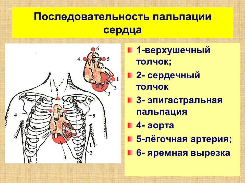 Palpation
 - Percussion of the heart. This method of studying the cardiovascular system makes it possible to approximately determine the size of the organ and its position in the chest by tapping. Due to this, a preliminary diagnosis can be made.
 Percussion
 - Auscultation of the heart. This is done using a stethoscope. This method consists of listening and allows you to assess the nature of the heart murmur and the deviation of the sound picture from the norm. The procedure must be carried out in complete silence.
 Auscultation
 - Blood pressure measurement. In this case, any type of tonometer is used that measures pressure to determine hypertension or hypotension.
The described methods of studying the cardiovascular system are carried out only during the initial examination; if the doctor finds certain deviations from the norm, he prescribes additional methods to check the activity of the heart, in this case it is used full examination with special equipment.
Electrocardiography
This diagnostic method allows you to record and then study the electrical impulses that the heart muscle produces during operation. If the heart is without pathologies, then electrical excitation passes through different parts of the heart with a certain sequence. If the excitability of the heart muscle fails, this indicates pathologies and possible diseases.
When the myocardium contracts and relaxes, all data is recorded and written in the form of teeth, after which the doctor receives a curve or graph.
 ECG curve
 The data is recorded by a special device called an electrocardiograph. This diagnostic method allows you to evaluate the frequency and uniformity of the heart rhythm, various electrical processes occurring in the organ. An ECG is performed to detect arrhythmia, ischemia, and heart attack.
Important! Shifts in the ECG curve arise not only due to malfunction hearts. The reason may be diseases not related to this organ: pneumonia, pleurisy, obesity, etc.
Electrocardiography may be included in comprehensive examination heart along with other methods.
In addition to taking a cardiogram at rest, other ECG techniques are used:
- Holter monitoring;
- bicycle ergometry.
In the first case, the study continues for a day. Equipment and sensors are connected to the patient, after which round-the-clock recording of indicators of changes in excitability begins. Often, this method is used for severe patients, or if the problem appears periodically, for example, with short-term arrhythmia.
In the second case, an ECG is taken before and after stress on the body. This method allows us to identify the patient’s susceptibility to physical activity. Bicycle ergometry is often used for ischemia, namely exertional angina.
Phonocardiography and echocardiography
Phonocardiography allows you to record all sounds and murmurs of the heart. Recording is performed through a phonocardiograph, which is usually an additional device to an electrocardiograph. This method of instrumental diagnosis allows you to evaluate the symptoms of diseases by sound.
 Phonocardiography
 Echocardiography is performed using ultrasound. Today there are several methods for conducting echocardiography:
- One-dimensional echocardiography allows you to obtain a projection of the organ in the plane. The heart is examined using this method to determine the thickness of the walls and the size of the cavities. Additionally, the functioning of the valves and the condition of the organ during and after contraction are assessed.
- Two-dimensional echocardiography provides a three-dimensional image of the organ being examined, so the method is considered more informative.
- Doppler echocardiography – diagnostics of blood flow inside the heart, used to assess hemodynamics, identify valve and septal defects, and the presence of shunts.
Radiography
Methods for studying the heart and blood vessels using X-rays allow us to evaluate the size and shape of the heart, large vessels, and the volume of fluid in the pericardial part. When using this method, a person receives a dose of radiation, so there is no point in using it unnecessarily. It is used when other methods do not provide adequate information about the condition of a person and his organ.
X-rays cannot be used to examine pregnant women. One of the types of radiography is tomography. The latter method is more informative, since the picture is displayed on the monitor screen, simulating the patient’s organ, however, the radiation exposure in this case is higher than with x-rays.
Radionuclide examination and angiocardiography
An isotope study of the heart, namely the radionuclide method, is carried out by introducing radioisotopes into the blood, which make it possible to further evaluate their distribution. This method helps determine the formation of blood clots in blood vessels, as well as myocardial infarction. In this case, the patient also receives radiation.
Angiocardiography involves injecting a radiopaque contrast agent directly into the heart. With its help, doctors can study many parameters of the heart chambers and blood vessels. A procedure is used to determine the feasibility of carrying out surgical intervention on the organ. This method is one of the main ones when examining for blood clots. Angiocardiography is performed by catheterization.
 Cardiac thrombosis
 Important! Only the doctor chooses how to check the blood vessels of the heart, by Dopplerography or angiography. The choice of method is influenced by many parameters, including the purpose of the study.
For each person and specific case, a certain type of diagnosis can be used, although in some situations more than one method may be used, but several at once. It depends on the state of health, the age of the patient and the reason why the heart hurts, that is, the existing pathology.
Home testing methods
You can check your heart function at home, and people over 40 years of age are recommended to do this more often in order to detect deterioration of the condition in time. For home diagnostics, a tonometer is used, which can measure blood pressure and pulse rate.
A tonometer can be used of any type, for which you have enough money. Measurements are taken only in a sitting or lying position, at rest. You can do them on both arms, but only on the elbow. If during measurements the pressure is more or less than 110/70-140/90 and the indicator remains long time, it is recommended to visit a cardiologist.
I work in a clinic, very often people come who are concerned about the state of their cardiovascular system and just want to check it. Basically they are afraid of having a heart attack and so on. Someone wants to be tested for sports, and there are people like that too.
First of all, this is electrocardiography. Such old method has not lost its relevance to this day, it is quite informative, but it does not provide comprehensive information, it has a certain diagnostic niche. A resting electrocardiogram is performed, the patient comes, lies down, electrodes are applied, and an electrocardiogram is taken. This is one of the main diagnostic methods.
The second routine diagnostic method is cardiac ultrasound or echocardiography. A routine echocardiographic examination is performed in a regular clinic. Because there are many different subtypes of echocardiography, but we do the main thing in the clinic; it exists in any medical institution where there are echocardiography machines and specialists. This is a very serious method that provides a lot of information.
Electrocardiography provides information about the electrical activity of the heart, and the heart has electrical activity. About how an impulse is conducted, and the heart is an impulse generator, it is a conducting system, so we monitor how impulses are generated, how they are conducted. There are disturbances in the generation of impulses, disturbances in the conduction of impulses and other disorders. These are all electrical disturbances of the heart, which are shown by a cardiogram.
A cardiogram can indirectly provide information that some chambers of the heart are dilated. That is, the left ventricle is dilated, left ventricular hypertrophy.
An electrocardiogram can reveal dangerous abnormalities; I can give an example from practice. A young patient came to us for a trivial reason, they took a cardiogram, and on the cardiogram they found signs of Brugada syndrome, which can at any time lead to sudden death. This dangerous syndrome can be easily identified without expensive research methods. The patient was sent to a specialized institution, where this diagnosis was confirmed, and the patient was installed with a cardioverter, a device that, when dangerous arrhythmias occur, stops it with an electric discharge, and the patient does not die.
Then an ultrasound examination of the heart. There are more in-depth research methods. I think it would be a good idea to have an electrocardiogram and ultrasound examination of the heart for all patients. Because ultrasound examination of the heart provides sufficient information about the size of the heart. Ultrasound provides very accurate information about the functioning of the heart valves; we can identify congenital and acquired heart defects. Some heart defects require urgent cardiac surgery. Both the cardiogram and the ultrasound method can detect scars due to heart attacks and other diseases. For example, from a cardiac ultrasound, we may suspect that a patient has increased pulmonary pressure. And pulmonary pressure can be increased with pulmonary embolism, and then the patient can simply be saved from death. These seem to be routine, but on the other hand, very important research methods.
I would like to emphasize that a doctor’s appointment begins with complaints; we evaluate the patient’s complaints. Complaints are quite a lot of information. Sometimes simply asking the patient can make a diagnosis. Subsequently, the doctor listens to the heart and lungs with a stethoscope, examines the patient, and this adds even more information.
I would like to give an interesting example. There was a patient who was admitted with complaints of shortness of breath and weakness. And the doctor who treated her heard a very rough noise from her. That is, during the examination, the doctor already suspected that something was wrong. She listened to the noise, suspected that there was, say, a heart defect, and referred her to an ultrasound specialist. The ultrasound doctor, having done the examination, wrote that everything was normal.
This means that an imbalance has arisen as a result. The doctor hears a very rough noise, suspects a heart defect, but in this particular case they suspected aortic stenosis, aortic valve stenosis. Having done research, aortic stenosis was not found, then the patient was discharged with treatment recommendations, with a diagnosis of aortic stenosis, the doctor decided not to cancel her concept. Despite the fact that ultrasound did not confirm this diagnosis, the patient was discharged with a diagnosis of aortic stenosis. But the condition became worse, shortness of breath increased, weakness progressed, and the patient sought an appointment at our clinic. The doctor listens to the murmur, we repeat the ultrasound of the heart, and on the ultrasound of the heart I see that the patient has a congenital heart defect. Moreover, this is quite obvious, and the defect is very serious; it requires urgent surgical intervention. This suggests that there are discrepancies.
Sometimes it is possible to diagnose a congenital defect based on such noise, because there are certain guidelines and rules, and you can still make a diagnosis without ultrasound. Of course, it is better to use such routine diagnostic methods at an appointment. Firstly, these methods are very simple, you can use them and immediately make a fairly accurate diagnosis. Ideally, the cardiologist himself should do this and be able to interpret the results of the methods.
It is also necessary to conduct ultrasound examination of blood vessels. For example, vessels of the neck, brachiocephalic arteries or vessels of other regions where they are clearly visible, any arteries. Usually they do the arteries of the legs, because these vessels do not lie deep, their walls are clearly visible, they are large in diameter, and the process of atherosclerosis is visible.